

Mechanisms of methylmercury-induced neurotoxicity: evidence from experimental studies
- PMID: 21683713
- PMCID: PMC3183295
- DOI: 10.1016/j.lfs.2011.05.019
Mechanisms of methylmercury-induced neurotoxicity: evidence from experimental studies
Abstract
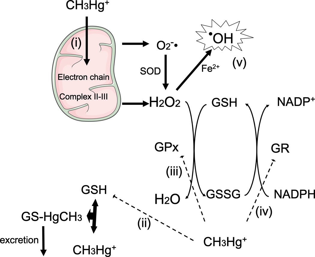
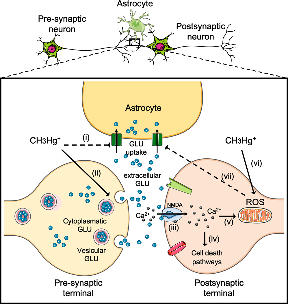
Neurological disorders are common, costly, and can cause enduring disability. Although mostly unknown, a few environmental toxicants are recognized causes of neurological disorders and subclinical brain dysfunction. One of the best known neurotoxins is methylmercury (MeHg), a ubiquitous environmental toxicant that leads to long-lasting neurological and developmental deficits in animals and humans. In the aquatic environment, MeHg is accumulated in fish, which represent a major source of human exposure. Although several episodes of MeHg poisoning have contributed to the understanding of the clinical symptoms and histological changes elicited by this neurotoxicant in humans, experimental studies have been pivotal in elucidating the molecular mechanisms that mediate MeHg-induced neurotoxicity. The objective of this mini-review is to summarize data from experimental studies on molecular mechanisms of MeHg-induced neurotoxicity. While the full picture has yet to be unmasked, in vitro approaches based on cultured cells, isolated mitochondria and tissue slices, as well as in vivo studies based mainly on the use of rodents, point to impairment in intracellular calcium homeostasis, alteration of glutamate homeostasis and oxidative stress as important events in MeHg-induced neurotoxicity. The potential relationship among these events is discussed, with particular emphasis on the neurotoxic cycle triggered by MeHg-induced excitotoxicity and oxidative stress. The particular sensitivity of the developing brain to MeHg toxicity, the critical role of selenoproteins and the potential protective role of selenocompounds are also discussed. These concepts provide the biochemical bases to the understanding of MeHg neurotoxicity, contributing to the discovery of endogenous and exogenous molecules that counteract such toxicity and provide efficacious means for ablating this vicious cycle.
Copyright © 2011 Elsevier Inc. All rights reserved.
Figures
Similar articles
-
Oxidative stress in MeHg-induced neurotoxicity.Toxicol Appl Pharmacol. 2011 Nov 1;256(3):405-17. doi: 10.1016/j.taap.2011.05.001. Epub 2011 May 9. Toxicol Appl Pharmacol. 2011. PMID: 21601588 Free PMC article. Review.
-
Involvement of glutamate and reactive oxygen species in methylmercury neurotoxicity.Braz J Med Biol Res. 2007 Mar;40(3):285-91. doi: 10.1590/s0100-879x2007000300001. Braz J Med Biol Res. 2007. PMID: 17334523 Review.
-
Protective effects of MK-801 on methylmercury-induced neuronal injury in rat cerebral cortex: involvement of oxidative stress and glutamate metabolism dysfunction.Toxicology. 2012 Oct 28;300(3):112-20. doi: 10.1016/j.tox.2012.06.006. Epub 2012 Jun 18. Toxicology. 2012. PMID: 22722016
-
Methylmercury-induced alterations in astrocyte functions are attenuated by ebselen.Neurotoxicology. 2011 Jun;32(3):291-9. doi: 10.1016/j.neuro.2011.01.004. Epub 2011 Feb 15. Neurotoxicology. 2011. PMID: 21300091 Free PMC article.
-
Protective effects of memantine against methylmercury-induced glutamate dyshomeostasis and oxidative stress in rat cerebral cortex.Neurotox Res. 2013 Oct;24(3):320-37. doi: 10.1007/s12640-013-9386-3. Epub 2013 Mar 16. Neurotox Res. 2013. PMID: 23504438
Cited by
-
Neuroprotective effect of Tagara, an Ayurvedic drug against methyl mercury induced oxidative stress using rat brain mitochondrial fractions.BMC Complement Altern Med. 2015 Aug 12;15:268. doi: 10.1186/s12906-015-0793-2. BMC Complement Altern Med. 2015. PMID: 26264039 Free PMC article.
-
Cellular transport and homeostasis of essential and nonessential metals.Metallomics. 2012 Jul;4(7):593-605. doi: 10.1039/c2mt00185c. Epub 2012 Feb 15. Metallomics. 2012. PMID: 22337135 Free PMC article. Review.
-
Effects of Gintonin-Enriched Fraction on Methylmercury-Induced Neurotoxicity and Organ Methylmercury Elimination.Int J Environ Res Public Health. 2020 Jan 29;17(3):838. doi: 10.3390/ijerph17030838. Int J Environ Res Public Health. 2020. PMID: 32013120 Free PMC article.
-
The catecholaminergic neurotransmitter system in methylmercury-induced neurotoxicity.Adv Neurotoxicol. 2017;1:47-81. doi: 10.1016/bs.ant.2017.07.002. Epub 2017 Sep 1. Adv Neurotoxicol. 2017. PMID: 32346666 Free PMC article. No abstract available.
-
Dietary Intakes of EPA and DHA Omega-3 Fatty Acids among US Childbearing-Age and Pregnant Women: An Analysis of NHANES 2001-2014.Nutrients. 2018 Mar 28;10(4):416. doi: 10.3390/nu10040416. Nutrients. 2018. PMID: 29597261 Free PMC article.
References
-
- Allen JW, Mutkus LA, Aschner M. Methylmercury-mediated inhibition of 3H-D-aspartate transport in cultured astrocytes is reversed by the antioxidant catalase. Brain Res. 2001;902(1):92–100. - PubMed
-
- Amonpatumrat S, Sakurai H, Wiriyasermkul P, Khunweeraphong N, Nagamori S, Tanaka H, Piyachaturawat P, Kanai Y. L-glutamate enhances methylmercury toxicity by synergistically increasing oxidative stress. J Pharmacol Sci. 2008;108(3):280–289. - PubMed
-
- Anderson CM, Swanson RA. Astrocyte glutamate transport: review of properties, regulation, and physiological functions. Glia. 2000;32(1):1–14. - PubMed
-
- Aschner M, Clarkson TW. Mercury 203 distribution in pregnant and nonpregnant rats following systemic infusions with thiol-containing amino acids. Teratology. 1987;36(3):321–328. - PubMed
-
- Aschner M, Syversen T, Souza DO, Rocha JB, Farina M. Involvement of glutamate and reactive oxygen species in methylmercury neurotoxicity. Braz J Med Biol Res. 2007;40(3):285–291. - PubMed
Publication types
MeSH terms
Substances
Grants and funding
LinkOut - more resources
Full Text Sources
