

Macroscopic and histopathologic analysis of human knee menisci in aging and osteoarthritis
- PMID: 21683797
- PMCID: PMC3217905
- DOI: 10.1016/j.joca.2011.05.008
Macroscopic and histopathologic analysis of human knee menisci in aging and osteoarthritis
Abstract
Objective: Meniscus lesions following trauma or associated with osteoarthritis (OA) have been described, yet meniscus aging has not been systematically analyzed. The objectives of this study were to (1) establish standardized protocols for representative macroscopic and microscopic analysis, (2) improve existing scoring systems, and (3) apply these techniques to a large number of human menisci.
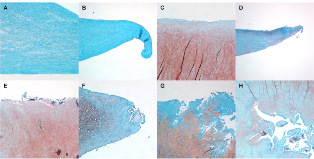
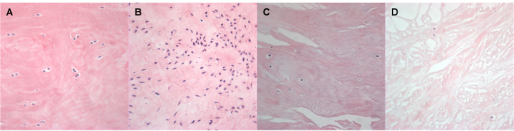
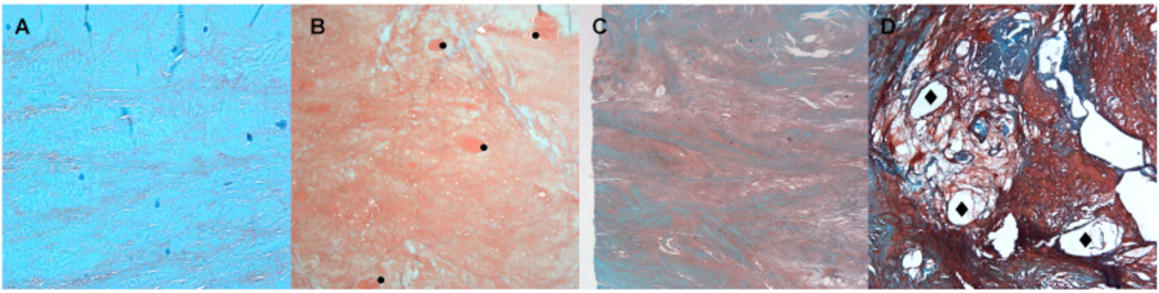
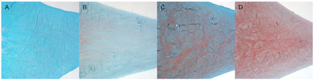
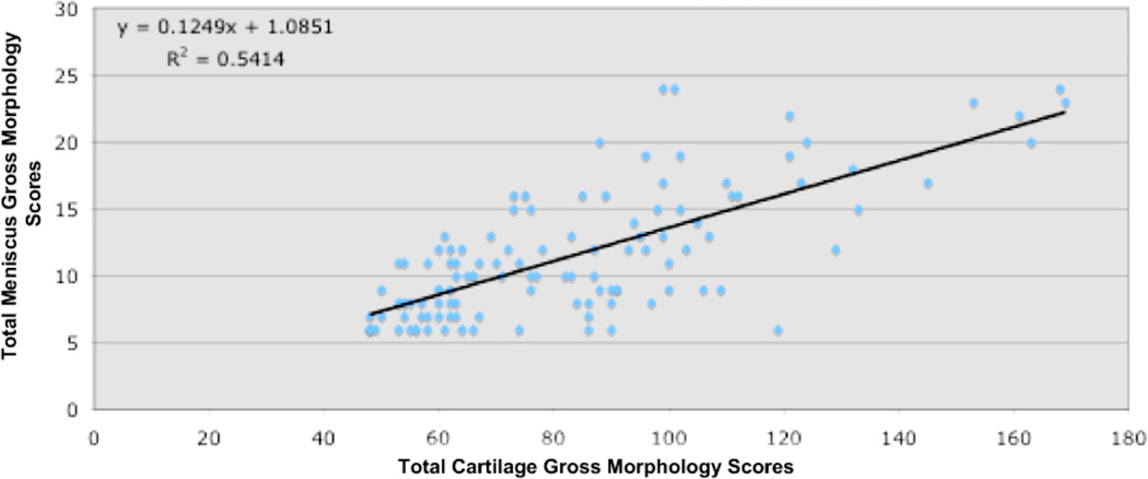
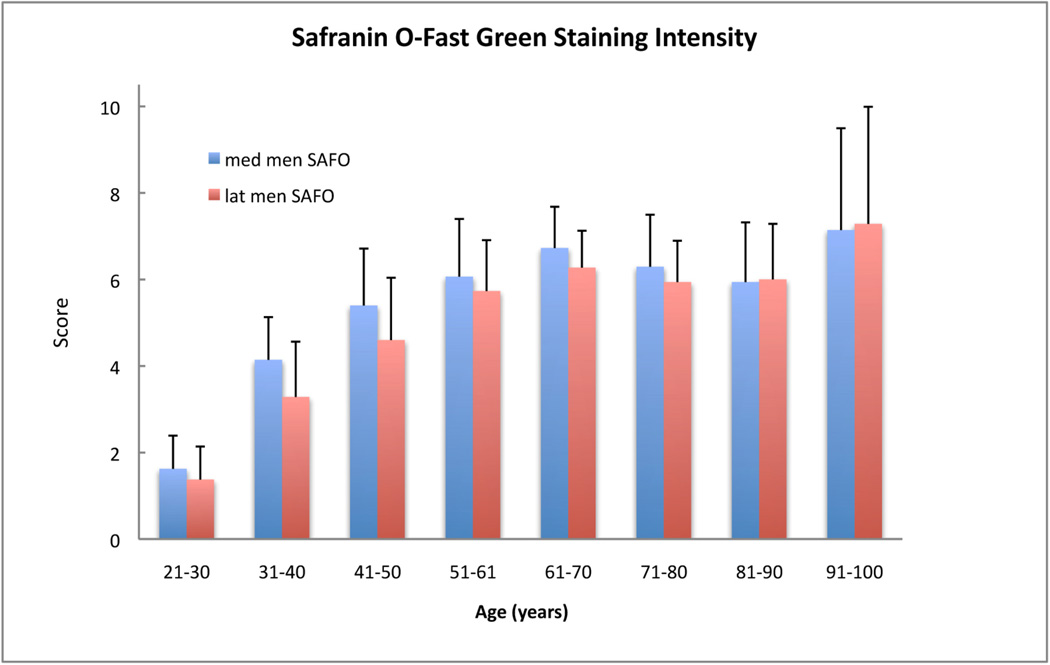
Design: Medial and lateral menisci from 107 human knees were obtained and cut in two different planes (triangle/cross section and transverse/horizontal section as well) in three separate locations (middle portion, anterior and posterior horns). All sections included vascular and avascular regions and were graded for (1) surface integrity, (2) cellularity, (3) matrix/fiber organization and collagen alignment, and (4) Safranin-O staining intensity. The cartilage in all knee compartments was also scored.
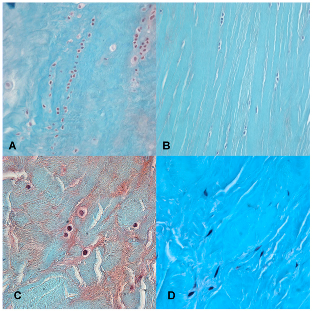
Results: The new macroscopic and microscopic grading systems showed high inter-reader and intra-reader intraclass correlation coefficients. The major age-related changes in menisci in joints with no or minimal OA included increased Safranin-O staining intensity, decreased cell density, the appearance of acellular zones, and evidence of mucoid degeneration with some loss of collagen fiber organization. The earliest meniscus changes occurred predominantly along the inner rim. Menisci from OA joints showed severe fibrocartilaginous separation of the matrix, extensive fraying, tears and calcification. Abnormal cell arrangements included decreased cellularity, diffuse hypercellularity along with cellular hypertrophy and abnormal cell clusters. In general, the anterior horns of both medial and lateral menisci were less affected by age and OA.
Conclusions: New standardized protocols and new validated grading systems allowed us to conduct a more systematic evaluation of changes in aging and OA menisci at a macroscopic and microscopic level. Several meniscus abnormalities appear to be specific to aging in the absence of significant OA. With aging the meniscal surface can be intact but abnormal matrix organization and cellularity were observed within the meniscal substance. The increased Safranin-O staining appears to represent a shift from fibroblastic to chondrocytic phenotype during aging and early degeneration.
Copyright © 2011 Osteoarthritis Research Society International. Published by Elsevier Ltd. All rights reserved.
Conflict of interest statement
No author has any conflict of interest related to this work.
Figures


References
-
- Song Y, Greve JM, Carter DR, Giori NJ. Meniscectomy alters the dynamic deformational behavior and cumulative strain of tibial articular cartilage in knee joints subjected to cyclic loads. Osteoarthritis Cartilage. 2008;16:1545–1554. - PubMed
-
- Seedholm B. Part II. Transmission of the load in the knee with special reference to the role of the meniscus. Eng Med. 1979;8:221–228.
-
- Seedholm B. Part I. Transmission of the load in the knee with special reference to the role of the meniscus. Eng Med. 1979;9:207–221.
-
- Lohmander LS, Englund PM, Dahl LL, Roos EM. The long-term consequence of anterior cruciate ligament and meniscus injuries: osteoarthritis. Am J Sports Med. 2007;35:1756–1769. - PubMed
-
- Gale DR, Chaisson CE, Totterman SM, Schwartz RK, Gale ME, Felson D. Meniscal subluxation: association with osteoarthritis and joint space narrowing. Osteoarthritis Cartilage. 1999;7:526–532. - PubMed
Publication types
MeSH terms
Grants and funding
LinkOut - more resources
Full Text Sources
Other Literature Sources
