. 2011 Nov 1;256(3):219-26.
doi: 10.1016/j.taap.2011.06.001. Epub 2011 Jun 13.

Protective effects of antioxidants and anti-inflammatory agents against manganese-induced oxidative damage and neuronal injury

Affiliations

Protective effects of antioxidants and anti-inflammatory agents against manganese-induced oxidative damage and neuronal injury

Dejan Milatovic et al. Toxicol Appl Pharmacol. .

Abstract

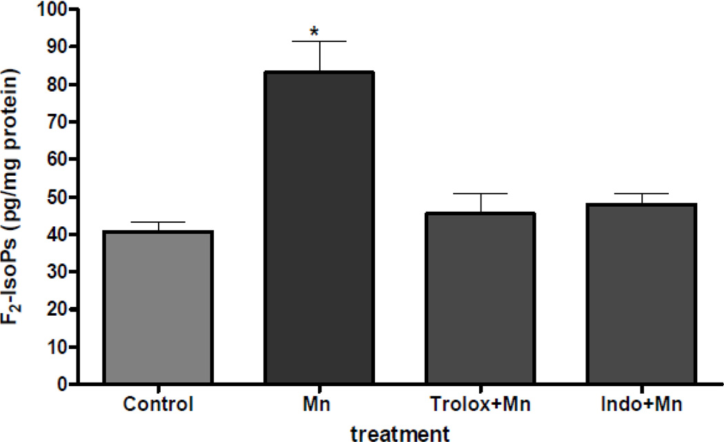
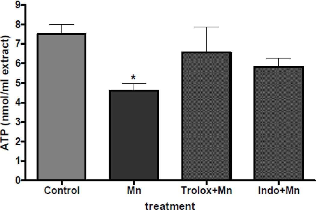
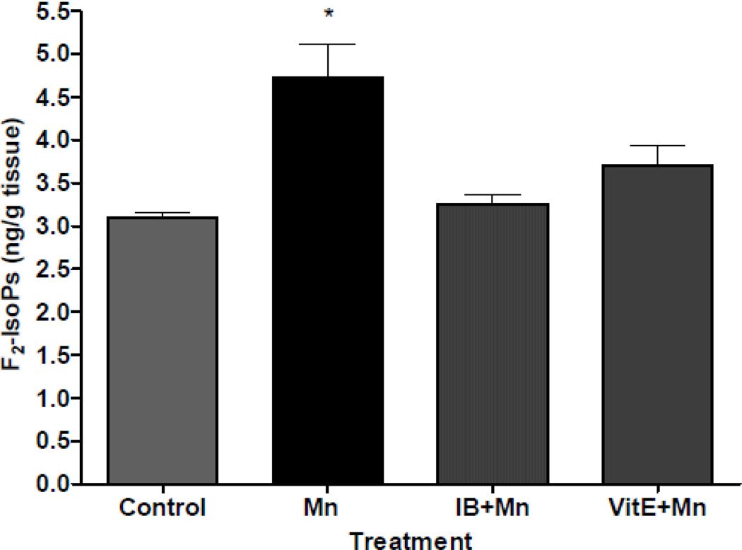
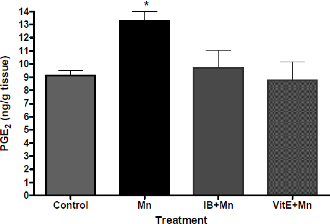
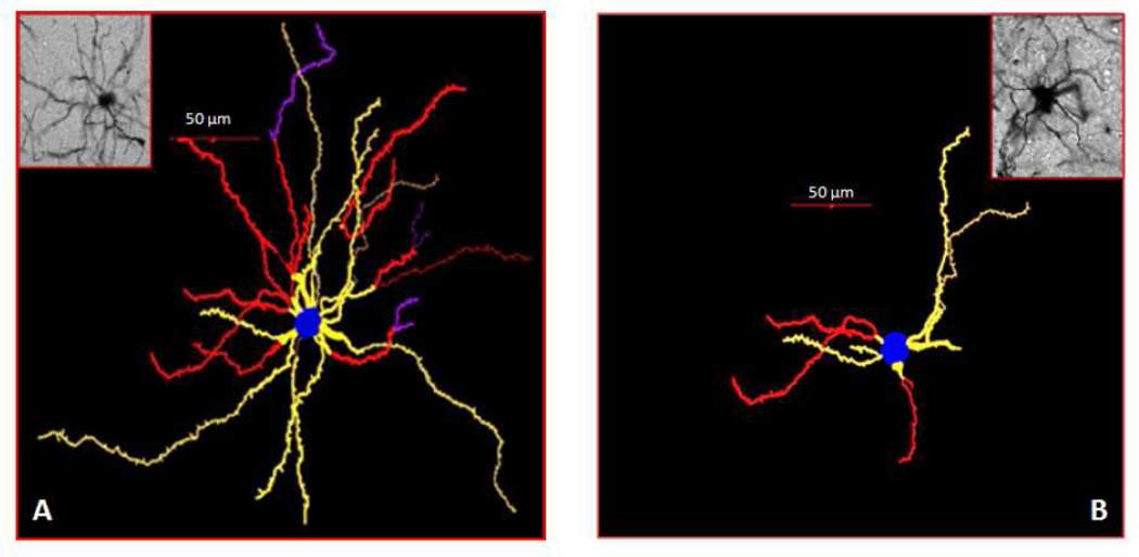
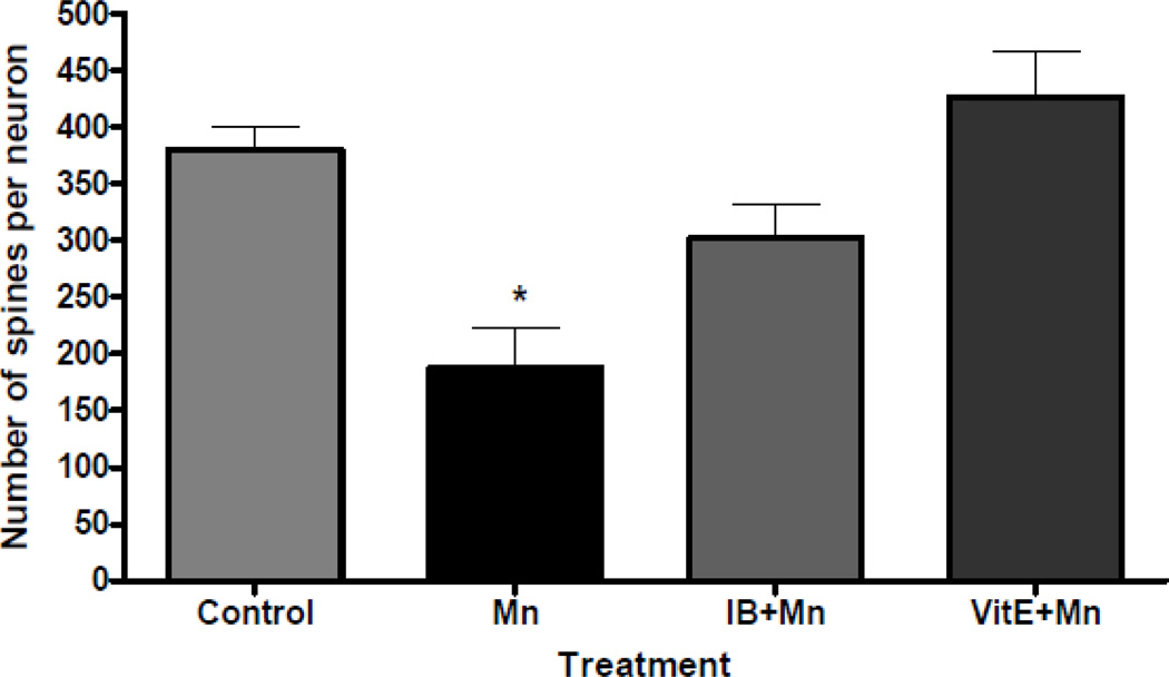
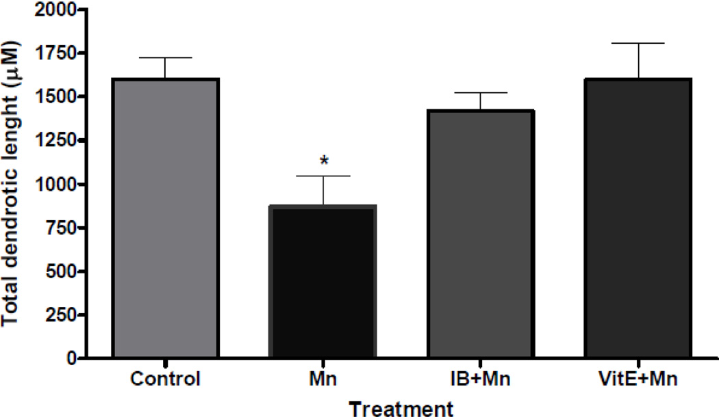
Exposure to excessive manganese (Mn) levels leads to neurotoxicity, referred to as manganism, which resembles Parkinson's disease (PD). Manganism is caused by neuronal injury in both cortical and subcortical regions, particularly in the basal ganglia. The basis for the selective neurotoxicity of Mn is not yet fully understood. However, several studies suggest that oxidative damage and inflammatory processes play prominent roles in the degeneration of dopamine-containing neurons. In the present study, we assessed the effects of Mn on reactive oxygen species (ROS) formation, changes in high-energy phosphates and associated neuronal dysfunctions both in vitro and in vivo. Results from our in vitro study showed a significant (p<0.01) increase in biomarkers of oxidative damage, F(2)-isoprostanes (F(2)-IsoPs), as well as the depletion of ATP in primary rat cortical neurons following exposure to Mn (500 μM) for 2h. These effects were protected when neurons were pretreated for 30 min with 100 of an antioxidant, the hydrophilic vitamin E analog, trolox (6-hydroxy-2,5,7,8-tetramethylchroman-2-carboxylic acid), or an anti-inflammatory agent, indomethacin. Results from our in vivo study confirmed a significant increase in F(2)-IsoPs levels in conjunction with the progressive spine degeneration and dendritic damage of the striatal medium spiny neurons (MSNs) of mice exposed to Mn (100mg/kg, s.c.) 24h. Additionally, pretreatment with vitamin E (100mg/kg, i.p.) or ibuprofen (140 μg/ml in the drinking water for two weeks) attenuated the Mn-induced increase in cerebral F(2)-IsoPs? and protected the MSNs from dendritic atrophy and dendritic spine loss. Our findings suggest that the mediation of oxidative stress/mitochondrial dysfunction and the control of alterations in biomarkers of oxidative injury, neuroinflammation and synaptodendritic degeneration may provide an effective, multi-pronged therapeutic strategy for protecting dysfunctional dopaminergic transmission and slowing of the progression of Mn-induced neurodegenerative processes.


Figures

Figure 1
Figure 1
Protection by trolox and indomethacin of Mn-induced changes in F2-IsoPs formation. Rat primary neuronal cultures were incubated with trolox or indomethacin (100 µM) for 30 min at 37°C, and F2-IsoPs levels were quantified at 2 hours following exposure to MnCl2 (500 µM). Data represent the mean ± S.E.M. from three independent experiments. * p<0.01 versus control by one-way ANOVA followed by Bonferroni’s multiple comparison tests.
Figure 2
Figure 2
Protection by trolox and indomethacin of Mn-induced changes in ATP formation. Rat primary neuronal cultures were incubated with trolox or indomethacin (100 µM) for 30 min at 37°C, and ATP levels were quantified at 2 hours following exposure to MnCl2 (500 µM). Data represent the mean ± S.E.M. from three independent experiments. * p<0.01 versus control by one-way ANOVA followed by Bonferroni’s multiple comparison tests.
Figure 3
Figure 3
Cerebral F2-IsoPs concentrations following saline- (control) or Mn- (100 mg/kg, s.c.) exposed mice with or without pretreatment with ibuprofen (IB, 140 µg/ml in drinking water for 2 weeks) or vitamin E (Vit E, α-tocopherol, 100 mg/kg, i.p., for 3 days). Brains from mice exposed to Mn were collected 24 hours post injections. Values of F2-IsoPs represent mean ± S.E.M. (n=4–6). *Significant difference between control mice and Mn-treated mice (p<0.05).
Figure 4
Figure 4
Cerebral PGE2 concentrations following saline- (control) or Mn- (100 mg/kg, s.c.) exposed mice with or without pretreatment with ibuprofen (IB, 140 µg/ml in drinking water for 2 weeks) or vitamin E (Vit E, α-tocopherol, 100 mg/kg, i.p., for 3 days). Brains from mice exposed to Mn were collected 24 hours post injections. Values of PGE2 represent mean ± S.E.M. (n=4–6). *Significant difference between control and Mn-treated mice (p<0.05).
Figure 5
Figure 5
Representative tracings of striatal medium spiny neurons (MSNs) with photomicrographs of mouse striatal sections from mice treated with saline (control) (A) or MnCl2 (100 mg/kg, s.c.) (B). The brain from mice exposed to MnCl2 was collected 24 hours after the injection. Treatment with Mn induced the degeneration of the striatal dendritic system as well as a decrease in the total number of spines and the length of the dendrites of MSNs. Tracing and counting were done using a Neurolucida system at 60× magnification (MicroBrightField, VT). Colors indicate the degree of dendritic branching (yellow=1°, red=2°, purple=3°).
Figure 6
Figure 6
Quantitative analyses of the total spine number of medium spiny neurons from the striatum of mice treated with saline (control) or Mn (100 mg/kg, s.c.) with or without pretreatment with ibuprofen (IB, 140 µg/ml in drinking water for 2 weeks) or vitamin E (Vit E, α-tocopherol, 100 mg/kg, i.p., for 3 days) and sacrificed 24 hours after the treatment. Tracing and counting of four to six Golgi-impregnated striatal MSNs were done using a Neurolucida system at 60× magnification (MicroBrightField, VT). *Significant difference between control and Mn-treated mice (P<0.05). Treatment with Mn induced the degeneration of the striatal dendritic system and a decrease in the total number of spines of MSNs.
Figure 7
Figure 7
Quantitative analyses of the total dendritic length of medium spiny neurons from the striatum of mice treated with saline (control) or Mn (100 mg/kg, s.c.) with or without pretreatment with ibuprofen (IB, 140 µg/ml in drinking water for 2 weeks) or viatmin E (Vit E, α-tocopherol, 100 mg/kg, i.p., for 3 days) and sacrificed 24 hours after the treatment. Tracing and counting of four to six Golgi-impregnated striatal MSNs were done using a Neurolucida system at 60× magnification (MicroBrightField, VT). *Significant difference between control and Mn-treated mice (P<0.05). Treatment with Mn induced the degeneration of the striatal dendritic system and as well as a decrease in the length of the dendrites of MSNs.

References

    1. Asanuma M, Nishibayashi-Asanuma S, Miyazaki I, Kohno M. Neuroprotective effects of non-steroidal anti-inflammatory drugs by direct scavenging of nitric oxide radicals. J. Neurochem. 2001;76:1895–1904. - PubMed
    1. Aschner M. Manganese in health and disease: from transport to neurotoxicity. In: Massaro E, editor. Handbook of neurotoxicology. Totowa, NJ: Humana Press; 2000. pp. 195–209.
    1. Autissier N, Rochette L, Dumas P, Beley A, Loireau A, Bralet J. Dopamine and norepinephrine turnover in various regions of the rat brain after chronic manganese chloride administration. Toxicology. 1982;24:175–182. - PubMed
    1. Avila DS, Colle D, Gubert P, Palma AS, Puntel G, Manarin F, Noremberg S, Nascimento PC, Aschner M, Rocha JB, Soares FA. A possible neuroprotective action of a vinylic telluride against Mn-induced neurotoxicity. Toxicol Sci. 2010;115:194–201. - PMC - PubMed
    1. Awas JA, Soteriou MC, Drougas JG, Stokes KA, Roberts LJ, 2nd, Pinson CW. Plasma prostaglandin E1 concentrations and hemodynamics during intravenous infusions of prostaglandin E1 in humans and swine. Transplantation. 1996;61:1624–1629. - PubMed

Publication types

MeSH terms