. 2011 Sep 8:190:228-38.
doi: 10.1016/j.neuroscience.2011.05.056. Epub 2011 Jun 13.

Change in number and activation of androgen receptor-immunoreactive cells in the medial amygdala in response to chemosensory input

Affiliations

Change in number and activation of androgen receptor-immunoreactive cells in the medial amygdala in response to chemosensory input

C B Blake et al. Neuroscience. .

Abstract

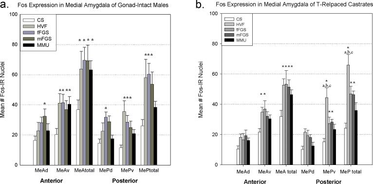
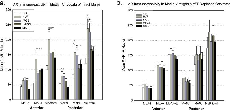
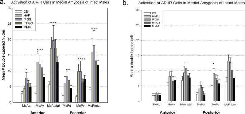
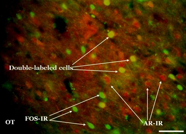
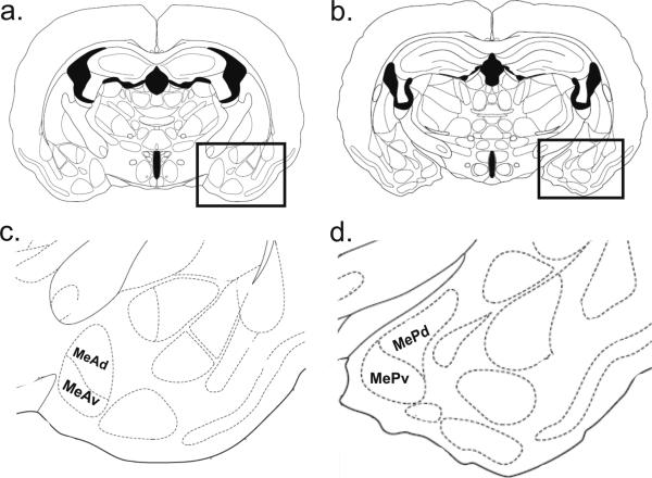
In many species social behaviors are dependent on integration of chemosensory and hormonal cues. Many chemosensory stimuli are detected by the vomeronasal organ, which projects to many regions that contain steroid receptors, including the medial amygdala. In male hamsters, testosterone is known to acutely increase in response to chemosensory stimulation, and can facilitate sexual behavior by direct action within the medial amygdala. Conspecific stimuli activate the anterior (MeA) and posterior (MeP) medial amygdala, while heterospecific stimuli activate only MeA. Chemosensory stimuli with different social significance differentially activate the dorsal and ventral subdivisions of MeA and MeP. Therefore, it is likely that steroids differentially facilitate stimulation of the medial amygdala by various chemosensory stimuli. We used Fos expression to examine activation of androgen receptor (AR)-containing cells in the medial amygdala by heterospecific and conspecific stimuli in intact male hamsters and castrated males with testosterone (T)-replacement. The number of AR-immunoreactive (-ir) cells was significantly different from control and between stimuli in intact males, but not in T-replaced castrates. Fos activation was similar in all animals. The results are consistent with a change in number of AR-ir cells in intact animals due to acute increases in testosterone caused by chemosignals.


Figures

Figure 1
Figure 1
Fos expression in the medial amygdala after exposure to various chemosensory stimuli in gonad-intact (a) and testosterone-replaced (b) male hamsters. Conspecific stimuli activated both anterior (MeA) and posterior (MeP) medial amygdala, while the heterospecific stimulus only activated MeA (a and b). Asterisk = significantly greater than control ( p<.03), a,b,c= significantly greater than MMU, fFGS, and mFGS respectively (p<.001 for all). CS= clean swab, HVF= hamster vaginal fluid, fFGS= female flank gland secretion, mFGS= male flank gland secretion, MMU= male mouse urine, d= dorsal subnucleus, v=ventral subnucleus.
Figure 2
Figure 2
Androgen receptor (AR)-immunoreactivity (ir) in the medial amygdala after exposure to various chemosensory stimuli gonad-intact (a) and testosterone-replaced (b) male hamsters. Conspecific and heterospecific stimuli significantly altered the number of AR-ir cells in different areas across the medial amygdala in gonad-intact animals (a). Conversely, there were no significant changes in testosterone-replaced castrates (b). Asterisk = significantly greater than control ( p<.05), a,b,c= significantly greater than MMU, fFGS, and mFGS respectively (p<.001 for all). CS= clean swab, HVF= hamster vaginal fluid, fFGS= female flank gland secretion, mFGS= male flank gland secretion, MMU= male mouse urine, MeA= anterior medial amygdala, MeP= posterior medial amygdala, d= dorsal subnucleus, v=ventral subnucleus.
Figure 3
Figure 3
Double-labeling of Fos and androgen receptors (AR) in the medial amygdala after exposure to various chemosensory stimuli gonad-intact (a) and testosterone-replaced (b) male hamsters. In gonad-intact males, conspecific stimuli yielded significantly more double-labeled cells than control (CS, clean swab) across the medial amygdala. The heterospecific stimulus (MMU, male mouse urine) produced significantly more double-labeled cells in the ventral portion of the posterior medial amygdala (MePv). Asterisk = significantly greater than control ( p<.05), a= significantly greater than MMU (p<.05). HVF= hamster vaginal fluid, fFGS= female flank gland secretion, mFGS= male flank gland secretion, MeA= anterior medial amygdala, MeP= posterior medial amygdala, d= dorsal subnucleus, v=ventral subnucleus.
Figure 4
Figure 4
Fos and androgen receptor (AR) double-label sample photomicrograph. Fos and ARs were labeled with monoclonal primary antibodies which were then recognized by fluorescent secondary antibodies. This photomicrograph was taken at the level of the posterior medial amygdala. This image was captured at 20x by a high-resolution black and white camera, and then pseudo-colored (Fos = green, AR = red) using Metamorph software. Scale bar = 100μm.
Figure 5
Figure 5
Schematic of anterior and posterior medial amygdala areas of interest. Coronal sections from the atlas of Morin and Wood (2001) at the level of anterior (a; Bregma: -1.2 mm.) and posterior (b; Bregma: -1.5 mm.) medial amygdala (boxed areas). The borders of the amygdala sub-regions used for counting are displayed in c and d (enlargements of the boxed regions of a and b, respectively).

References

    1. Bartke A. Male reproductive endocrinology. In: Siegel Hl., editor. The hamster: reproduction and behavior. Plenum Press; New York: 1985.
    1. Baum MJ, Everitt BJ. Increased expression of c-fos in the medial preoptic area after mating in male rats: role of afferent inputs from the medial amygdala and midbrain central tegmental field. Neurosci. 1992;50:627–646. - PubMed
    1. Bisler S, Schleicher A, Gass P, Stehle G, Zilles K, Staiger JF. Expression of c-Fos, ICER, Krox-24 and Jun B in the whisker-to-barrel pathway of rats: Time course of induction upon whisker stimulation by tactile exploration of an enriched environment. J Chemical Neuroanat. 2002;23:187–198. - PubMed
    1. Blake CB, Meredith M. Selective enhancement of main olfactory input to the medial amygdala by GnRH. Brain Res. 2010;1317:46–59. - PMC - PubMed
    1. Canteras NS. The medial hypothalamic defensive system: hodological organization and functional implications. Pharmacol Biochem Behav. 2002;71:481–491. - PubMed

Publication types