. 2011 Oct;44(1):19-27.
doi: 10.1016/j.nbd.2011.05.019. Epub 2011 Jun 6.

The role of CXCR4 signaling in the migration of transplanted oligodendrocyte progenitors into the cerebral white matter

Affiliations

The role of CXCR4 signaling in the migration of transplanted oligodendrocyte progenitors into the cerebral white matter

Ghazal Banisadr et al. Neurobiol Dis. 2011 Oct.

Abstract

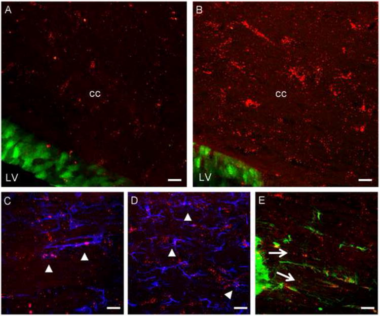
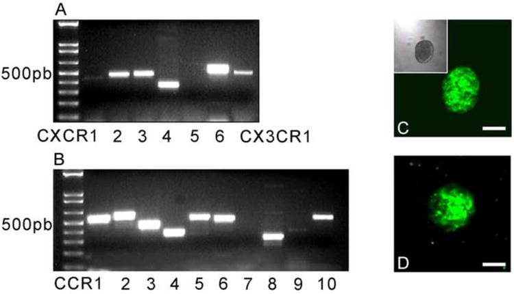
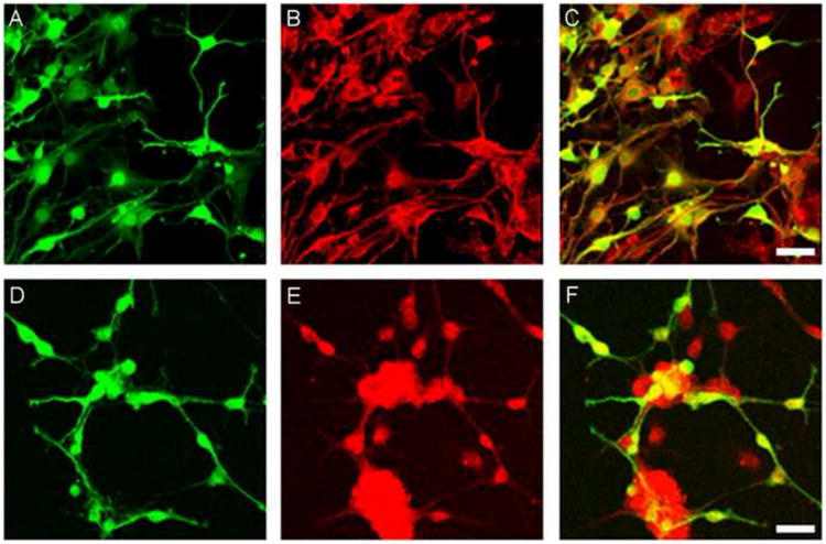
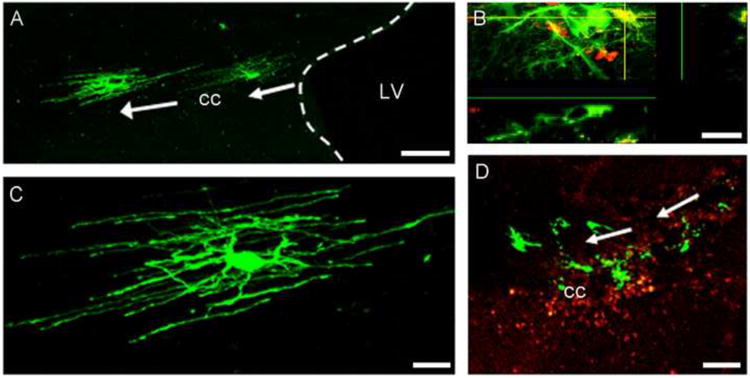
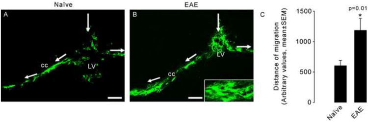
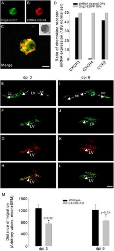
Enhancing the ability of either endogenous or transplanted oligodendrocyte progenitors (OPs) to engage in myelination may constitute a novel therapeutic approach to demyelinating diseases of the brain. It is known that in adults neural progenitors situated in the subventricular zone of the lateral ventricle (SVZ) are capable of generating OPs which can migrate into white matter tracts such as the corpus callosum (CC). We observed that progenitor cells in the SVZ of adult mice expressed CXCR4 chemokine receptors and that the chemokine SDF-1/CXCL12 was expressed in the CC. We therefore investigated the role of chemokine signaling in regulating the migration of OPs into the CC following their transplantation into the lateral ventricle. We established OP cell cultures from Olig2-EGFP mouse brains. These cells expressed a variety of chemokine receptors, including CXCR4 receptors. Olig2-EGFP OPs differentiated into CNPase-expressing oligodendrocytes in culture. To study the migratory capacity of Olig2-EGFP OPs in vivo, we transplanted them into the lateral ventricles of mice. Donor cells migrated into the CC and differentiated into mature oligodendrocytes. This migration was enhanced in animals with Experimental Autoimmune Encephalomyelitis (EAE). Inhibition of CXCR4 receptor expression in OPs using shRNA inhibited the migration of transplanted OPs into the white matter suggesting that their directed migration is regulated by CXCR4 signaling. These findings indicate that CXCR4 mediated signaling is important in guiding the migration of transplanted OPs in the context of inflammatory demyelinating brain disease.


Figures

Figure 1
Figure 1. Expression of SDF-1-RFP and CXCR4-EGFP in SDF-1-RFP/CXCR4-EGFP bitransgenic mouse with EAE
SDF-1-RFP/CXCR4-EGFP bitransgenic mice illustrate the presence of SDF-1-RFP (red) in the corpus callosum (cc) and CXCR4-EGFP (green) in the wall of the lateral ventricle (LV) in naïve (A) and EAE (B) mice brains. The expression of SDF-1 in the CC was greatly upregulated in mice with EAE (B) and was expressed by GFAP-labeled astrocytes (blue) and IBA-1- labeled microglia (blue) as shown in panels C and D respectively (arrowheads). CXCR4 is expressed by cells showing the morphology of migrating progenitors in the posterior part of the SVZ (E). Scale bars=50μm.
Figure 2
Figure 2. Expression of chemokine receptors by oligodendrocyte progenitor cells
Total RNA was extracted from oligospheres and RT-PCR was performed. Chemokine receptors of the CXC family (A), CCR family (B) and the chemokine receptor CX3CR1 (A) were expressed by OP cells. Panels C and D show oligosphere cultures from Olig-2-EGFP and CXCR4-EGFP transgenic mice respectively. Scale bars=50μm.
Figure 3
Figure 3. Olig2-EGFP and CXCR4-EGFP oligospheres differentiate into oligodendrocytes in vitro
The ability of oligospheres to generate differentiated oligodendrocytes was examined in vitro following plating them on coverslips. Oligospheres from Olig2-EGFP and CXCR4-EGFP transgenic mice generated the morphology of oligodendrocytes (panels A and D respectively). Immunohistochemistry was performed using an oligodendrocyte marker, CNPase (red labeling in panels B and E). Panels C and F show the co-localization of Olig2-EGFP (C) and CXCR4-EGFP (F) oligodendrocytes with CNPase. Scale bars=50μm.
Figure 4
Figure 4. Transplanted Olig2-EGFP OPs integrate into the demyelinated white matter and differentiate into oligodendrocytes in vivo
Dissociated OPs from Olig2-EGFP mice were injected into the lateral ventricle (LV) of EAE mice at the peak of the disease. Two weeks post-transplantation, Olig2-EGFP cells migrated into the media-lateral axis of the corpus callosum (cc) (A) and differentiated into cells with the morphology of oligodendrocytes (C). Panel B shows the co-localization of a differentiated Olig2-EGFP cell (green) with the oligodendrocyte marker, CNPase (red). Panel D shows that Olig2-EGFP cells (green) migrate to areas of demyelination as characterized using an anti-PLP antibody (red) that stains for the myelin surrounding the demyelinated area. Scale bars in A and D=100μm, scale bars in C and B =20μm.
Figure 5
Figure 5. Olig2-EGFP OPs migrate further in EAE than in control mice
Olig2-EGFP OPs were injected into the lateral ventricle (LV) of naïve (A) and EAE (B) mice. Cells migrated into the corpus callosum (cc) and differentiated into oligodendrocytes (insert in B). Quantification of the migration distance showed that cells migrate further in EAE animals. As seen in panel B, more cells are observed in EAE tissue. The histogram in C shows the average distance of migration for oligospheres in naïve (n=6) and EAE (n=10) mice (t test, P=0.01). Scale bars=100μm.
Figure 6
Figure 6. Impaired migration of OPs in EAE mice following downregulation of CXCR4 expression
Olig2-EGFP oligospheres were infected with an adenovirus, which expresses shRNA targeting CXCR4 and a fluorescent reporter protein (DsRed2) (A-C). Histogram in D illustrates the ratio of chemokine receptor mRNA expression in shRNA treated OPs to non-infected Olig2-EGFP OPs. Quantitative PCR showed that the expression of CXCR4 was significantly downregulated in shRNA expressing cells while the expression of other chemokine receptors e.g. CXCR3 and CCR2 was not downregulated. Three days post injection (dpi), both untreated Olig2-EGFP (E) and shRNA treated cells (F-H) migrated into the corpus callosum (cc) and differentiated into oligodendrocytes. Green labeling shows the Olig-2-EGFP expression and the red fluorescence corresponds to DsRed. The merged cells expressing both green and red fluorescence (H) are cells infected with the shRNA (CXCR4 KO). At 6 days post injection, cells obtained from untreated Olig2-EGFP (I) and shRNA treated Olig2-EGFP OPs (J-L) were still observed in the corpus callosum of EAE mice. Migration distance in arbitrary units was calculated using the Metamorph program. Measurements were taken from the intersection of the injection path and the stem cell migratory stream to the most medially and most laterally migrated cell body. The data were reported as the mean±SEM of the total (medial + lateral) migration per experimental group. We quantified the differentiated cells by counting EGFP and/or DsRed2 positive cells within the corpus callosum and presented the data as mean±SEM per square millimeter of the white matter. Quantification of the lateral and medial migration of OPs showed that CXCR4 KO cells migrated less than untreated (wildtype) Olig2-EGFP cells (histogram in M) or cells treated with an adenovirus expressing DsRed2 but no shRNA (not shown in the histogram). Similar results were obtained at 3 and 6 days post injection. Asterisks mark statistically significant results (t test, p=0.02 and 0.03). Scale bars=100μm.

References

    1. Aboody KS, Brown A, Rainov NG, Bower KA, Liu S, Yang W, Small JE, Herrlinger U, Ourednik V, Black PM, Breakefield XO, Snyder EY. Neural stem cells display extensive tropism for pathology in adult brain: evidence from intracranial gliomas. Proc Natl Acad Sci U S A. 2000;97:12846–51. - PMC - PubMed
    1. Bagri A, Gurney T, He X, Zou YR, Littman DR, Tessier-Lavigne M, Pleasure SJ. The chemokine SDF1 regulates migration of dentate granule cells. Development. 2002;129:4249–60. - PubMed
    1. Banisadr G, Skrzydelski D, Kitabgi P, Rostene W, Parsadaniantz SM. Highly regionalized distribution of stromal cell-derived factor-1/CXCL12 in adult rat brain: constitutive expression in cholinergic, dopaminergic and vasopressinergic neurons. Eur J Neurosci. 2003;18:1593–606. - PubMed
    1. Belmadani A, Tran PB, Ren D, Assimacopoulos S, Grove EA, Miller RJ. The chemokine stromal cell-derived factor-1 regulates the migration of sensory neuron progenitors. J Neurosci. 2005;25:3995–4003. - PMC - PubMed
    1. Belmadani A, Tran PB, Ren D, Miller RJ. Chemokines regulate the migration of neural progenitors to sites of neuroinflammation. J Neurosci. 2006;26:3182–91. - PMC - PubMed

Publication types

MeSH terms