. 2011 Jun 17;18(7):435-43.
doi: 10.1101/lm.2132911. Print 2011 Jul.

Separate but interacting recognition memory systems for different senses: the role of the rat perirhinal cortex

Affiliations

Separate but interacting recognition memory systems for different senses: the role of the rat perirhinal cortex

Mathieu M Albasser et al. Learn Mem. .

Abstract

Two different models (convergent and parallel) potentially describe how recognition memory, the ability to detect the re-occurrence of a stimulus, is organized across different senses. To contrast these two models, rats with or without perirhinal cortex lesions were compared across various conditions that controlled available information from specific sensory modalities. Intact rats not only showed visual, tactile, and olfactory recognition, but also overcame changes in the types of sensory information available between object sampling and subsequent object recognition, e.g., between sampling in the light and recognition in the dark, or vice versa. Perirhinal lesions severely impaired object recognition whenever visual cues were available, but spared olfactory recognition and tactile-based object recognition when tested in the dark. The perirhinal lesions also blocked the ability to recognize an object sampled in the light and then tested for recognition in the dark, or vice versa. The findings reveal parallel recognition systems for different senses reliant on distinct brain areas, e.g., perirhinal cortex for vision, but also show that: (1) recognition memory for multisensory stimuli involves competition between sensory systems and (2) perirhinal cortex lesions produce a bias to rely on vision, despite the presence of intact recognition memory systems serving other senses.


Figures
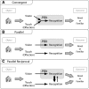
Figure 1.
Figure 1.
Schematic diagram showing hypothetical models of the interactions between sensory recognition systems. (A) Convergent: Perirhinal cortex receives information for recognition from different senses. (B) Parallel: Different sensory recognition systems work in parallel with the perirhinal cortex involved only in vision. For Model B it is assumed that the detection of novelty by any one stream is sufficient to make a “new” decision. (C) Parallel Reciprocal: The perirhinal cortex is vital for the visual system but also interacts with other sensory systems. For example, vision might override touch or olfaction (bold arrow between the two systems). Olfaction is assumed to mirror the organization for touch, and so is placed in parentheses. The converging arrows represent multisensory systems that are then unitized prior to making the decision whether the object is novel or familiar.
Figure 2.
Figure 2.
Experiment 1: Object recognition of junk objects in the light and dark by rats with perirhinal cortex lesions (black) and surgical controls (white). Rats with perirhinal lesions are particularly impaired when tested in the light. The histograms (left) show the D2 scores when the data from all 10 trials are considered; the graphs (right) show the updated D2 scores over successive trials. D2 is the time exploring the novel object minus the time exploring the familiar object, divided by total exploration. Scores can range from +1 to −1. Data shown are mean ± standard error of the mean. Group differences ***P < 0.001.
Figure 3.
Figure 3.
Experiment 2: Odor recognition in the light and dark by rats with perirhinal cortex lesions (black) and surgical controls (white). Rats with perirhinal lesions were not impaired, though neither group showed novel odor recognition in the light. The histograms (left) show the D2 scores when the data from all 10 trials are considered; the graphs (right) show the updated D2 scores over successive trials. D2 is the time exploring the novel object minus the time exploring the familiar object, divided by total exploration. Scores can range from +1 to −1. Data shown are mean ± standard error of the mean.
Figure 4.
Figure 4.
Experiment 3: Visual and shape recognition (Duplo objects) in the light and in the dark by rats with perirhinal cortex lesions (black) and surgical controls (white). Rats with perirhinal lesions were only impaired when tested in the light. The histograms (left) show the D2 scores when the data from all 10 trials are considered; the graphs (right) show the updated D2 scores over successive trials. D2 is the time exploring the novel object minus the time exploring the familiar object, divided by total exploration. Scores can range from +1 to −1. Data shown are mean ± standard error of the mean. Group differences ***P < 0.001.
Figure 5.
Figure 5.
Experiment 4: Object recognition—transfer between object sampling in the light or in the dark to a recognition test in the light or the dark. The resulting four conditions were: light to light (L → L), light to dark (L → D), dark to dark (D → D), and dark to light (D → L). Rats with perirhinal lesions (black histograms) were consistently impaired whenever sampled or tested in the light, but performed at normal levels for D → D. The histograms show the D2 scores when the data from all five trials at each condition are combined. D2 is the time exploring the novel object minus the time exploring the familiar object, divided by total exploration. Scores can range from +1 to −1. Data shown are mean ± standard error of the mean. Group differences ***P < 0.001.
Figure 6.
Figure 6.
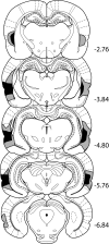
Diagrammatic reconstructions of the perirhinal cortex lesions showing the individual cases with the largest (gray) and smallest (black) lesions. The numbers refer to the distance (in millimeters) from bregma (adapted, with permission of Elsevier © 2005, from Paxinos and Watson 2005). The PRh group comprised 12 rats.
Figure 7.
Figure 7.
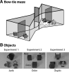
(A) Schematic of the bow-tie maze. A central sliding door separates the two ends of the maze in which two objects are placed. (B) Photographs showing examples of objects used in Experiment 1—junk objects (left); Experiment 2—odor cubes (middle; visually identical but with different aromas, e.g., rose, peach, or grass); Experiment 3—Duplo objects (right).

References

    1. Aggleton JP, Keen S, Warburton EC, Bussey TJ 1997. Extensive cytotoxic lesions of the rhinal cortices impair recognition but spare spatial alternation in the rat. Brain Res Bull 43: 279–287 - PubMed
    1. Albasser MM, Chapman RJ, Amin E, Iordanova MD, Vann SD, Aggleton JP 2010a. New behavioral protocols used to extend our knowledge of rodent object recognition memory. Learn Mem 17: 407–419 - PMC - PubMed
    1. Albasser MM, Poirier GL, Aggleton JP 2010b. Qualitatively different modes of perirhinal-hippocampal engagement when rats explore novel vs. familiar objects as revealed by c-fos imaging. Eur J Neurosci 31: 134–147 - PMC - PubMed
    1. Brown MW, Aggleton JP 2001. Recognition memory: What are the roles of the perirhinal cortex and hippocampus? Nat Rev Neurosci 2: 51–61 - PubMed
    1. Bucci DJ, Saddoris MP, Burwell RD 2002. Contextual fear discrimination is impaired by damage to the postrhinal or perirhinal cortex. Behav Neurosci 116: 479–488 - PubMed

Publication types

MeSH terms

Substances

LinkOut - more resources