. 2011 Aug;10(8):1095-9.
doi: 10.1128/EC.00002-11. Epub 2011 Jun 17.

Evidence for host cells as the major contributor of lipids in the intravacuolar network of Toxoplasma-infected cells

Affiliations

Evidence for host cells as the major contributor of lipids in the intravacuolar network of Toxoplasma-infected cells

Carolina E Caffaro et al. Eukaryot Cell. 2011 Aug.

Abstract

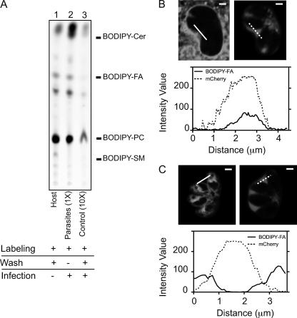
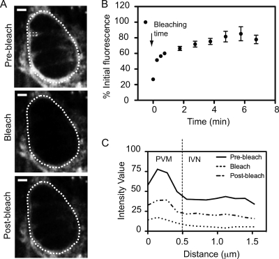
The intracellular parasite Toxoplasma gondii develops inside a parasitophorous vacuole (PV) that derives from the host cell plasma membrane during invasion. Previous electron micrograph images have shown that the membrane of this vacuole undergoes an extraordinary remodeling with an extensive network of thin tubules and vesicles, the intravacuolar network (IVN), which fills the lumen of the PV. While dense granule proteins, secreted during and after invasion, are the main factors for the organization and tubulation of the network, little is known about the source of lipids used for this remodeling. By selectively labeling host cell or parasite membranes, we uncovered evidence that strongly supports the host cell as the primary, if not exclusive, source of lipids for parasite IVN remodeling. Fluorescence recovery after photobleaching (FRAP) microscopy experiments revealed that lipids are surprisingly dynamic within the parasitophorous vacuole and are continuously exchanged or replenished by the host cell. The results presented here suggest a new model for development of the parasitophorous vacuole whereby the host provides a continuous stream of lipids to support the growth and maturation of the PVM and IVN.


Figures

Fig. 1.
Fig. 1.
Selective prelabeling of host or parasite membranes indicates that the host is the major lipid contributor to IVN development. (A) HFFs grown in 15-cm dishes were incubated with C4-BODIPY-C9-labeled fatty acid alone (lane 1; host), prelabeled with C4-BODIPY-C9-labeled fatty acid and infected with Toxoplasma for 18 h in the presence of the fluorescent probe [lane 2; parasites (1×)], or prelabeled with C4-BODIPY-C9-labeled fatty acid, washed, and infected with Toxoplasma for 18 h in medium without fluorescent probe [lane 3; control (10×)]. Lipids were extracted from host cells (lane 1) or purified parasites (lanes 2 and 3) and separated on silica plates by TLC using chloroform-methanol-water-triethylamine (35:30:7:35) as the solvent. For the control (10×), 10-fold more sample was spotted on the silica plate than for the parasite (1×). Fluorescence standards used were as follows: BODIPY-Cer, BODIPY-ceramide; BODIPY-FA, BODIPY fatty acid; BODIPY-PC, BODIPY-phosphatidylcholine; and BODIPY-SM, BODIPY-sphingomyelin. (B) HFFs grown on chambered cover glass were prelabeled with C4-BODIPY-C9-labeled fatty acid, washed, and infected with Toxoplasma secreting mCherry for 18 h. Optical sections (0.5 μm) of infected live cells were acquired by confocal microscopy. One section is shown. The left image represents the BODIPY label, and the right image shows secreted mCherry as a marker for the PV space. Intensity profiles are shown along the solid line for the BODIPY label and along the dashed line for mCherry, from left to right. Bars, 2 μm. (C) Same as panel B, except that parasites prelabeled with C4-BODIPY-C9-labeled fatty acid were used to infect unlabeled host cells.
Fig. 2.
Fig. 2.
Continuous flow of host lipids to Toxoplasma PVM and IVN. (A) Typical FRAP experiment with prelabeled HFFs infected with unlabeled parasites. A 1-μm optical slice was taken before bleaching (prebleach), right after the photobleach (bleach), and 2 min into recovery (postbleach), using an argon laser at 5% power. The white dotted line shows the perimeter of the area that was photobleached; the small dotted rectangle shows the portion of the IVN that was also analyzed in panel C. (B) Same as panel A, except that the argon laser was used at 2% power and the fluorescence intensity of the PV was normalized to prebleach values for prelabeled host cells infected with unlabeled parasites for 18 h and plotted over time. Error bars indicate standard deviations (n = 12 vacuoles). (C) The recovery of fluorescence within the PV and IVN after photobleaching of the full vacuole is shown by plotting of the intensity profile corresponding to the rectangular area indicated in panel A before, during, and 2 min after photobleaching. The dotted line at 0.5 μm represents the approximate boundary between the PVM at the periphery of the PV and the IVN occupying the space between the parasites.

Similar articles

Cited by

References

    1. Bai J., Pagano R. E. 1997. Measurement of spontaneous transfer and transbilayer movement of BODIPY-labeled lipids in lipid vesicles. Biochemistry 36:8840–8848 - PubMed
    1. Charron A. J., Sibley L. D. 2002. Host cells: mobilizable lipid resources for the intracellular parasite Toxoplasma gondii. J. Cell Sci. 115:3049–3059 - PubMed
    1. Coppens I., et al. 2006. Toxoplasma gondii sequesters lysosomes from mammalian hosts in the vacuolar space. Cell 125:261–274 - PubMed
    1. Coppens I., Sinai A. P., Joiner K. A. 2000. Toxoplasma gondii exploits host low-density lipoprotein receptor-mediated endocytosis for cholesterol acquisition. J. Cell Biol. 149:167–180 - PMC - PubMed
    1. Crawford M. J., et al. 2006. Toxoplasma gondii scavenges host-derived lipoic acid despite its de novo synthesis in the apicoplast. EMBO J. 25:3214–3222 - PMC - PubMed

Publication types