Comparative Study
. 2011 Jul 15;187(2):692-701.
doi: 10.4049/jimmunol.1003658. Epub 2011 Jun 17.

Notch signaling regulates mouse and human Th17 differentiation

Affiliations
Comparative Study

Notch signaling regulates mouse and human Th17 differentiation

Shilpa Keerthivasan et al. J Immunol. .

Abstract

Th17 cells are known to play a critical role in adaptive immune responses to several important extracellular pathogens. Additionally, Th17 cells are implicated in the pathogenesis of several autoimmune and inflammatory disorders as well as in cancer. Therefore, it is essential to understand the mechanisms that regulate Th17 differentiation. Notch signaling is known to be important at several stages of T cell development and differentiation. In this study, we report that Notch1 is activated in both mouse and human in vitro-polarized Th17 cells and that blockade of Notch signaling significantly downregulates the production of Th17-associated cytokines, suggesting an intrinsic requirement for Notch during Th17 differentiation in both species. We also present evidence, using promoter reporter assays, knockdown studies, as well as chromatin immunoprecipitation, that IL-17 and retinoic acid-related orphan receptor γt are direct transcriptional targets of Notch signaling in Th17 cells. Finally, in vivo inhibition of Notch signaling reduced IL-17 production and Th17-mediated disease progression in experimental autoimmune encephalomyelitis, a mouse model of multiple sclerosis. Thus, this study highlights the importance of Notch signaling in Th17 differentiation and indicates that selective targeted therapy against Notch may be an important tool to treat autoimmune disorders, including multiple sclerosis.


Figures
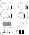
FIGURE 1
FIGURE 1
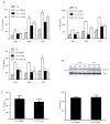
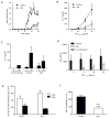
Gamma secretase inhibitors (GSI) significantly down-regulate Th17 associated cytokine levels in murine Th17 in-vitro polarization assays. (A) ELISA for IL-17A, IL-17F and IL-21 in supernatants of activated CD4+ T cells from C57BL/6 mice. Cells were pretreated in-vitro with GSI (25μM ILCHO and 4μM Compound E) or with 0.1% DMSO (as a vehicle control) before 24, 48 and 72 hours culture in Th17 polarizing conditions. Cells were then lifted, recounted and cultured overnight. (B) Notch1 expression in cells pretreated with or without ILCHO was evaluated by immunoblotting using antibodies that recognized the cleaved active Notch1. Antibody specific for actin was used to control for loading. (C) Evaluation of Notch1 expression in cells pretreated with or without ILCHO by flow cytometry using antibodies specific for CD4+ cells and intracellular Notch 1 (Notch1IC). (D) Intracellular staining of IL-17 and IFNγ in Th17 differentiated cells treated with either DMSO or GSI. (E) Naïve CD4+ T cells were differentiated towads Th17 subset for 4 days followed by treatment with either DMSO or GSI. Supernatants were collected after 24 h and IL-17 A ELISA was performed. Data shown here represents one of at least three independent experiments done in triplicates.*p≤0.05, **p≤0.001, ***p≤0.0001.
FIGURE 2
FIGURE 2
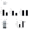
Gamma secretase inhibitors (GSIs) significantly reduce Th17 cytokine levels in human in-vitro Th17 polarization assays. (A) ELISA of IL-17A, IL-17F and IL-22 in supernatants of Th17 polarized naïve human CD4+ T cells treated with GSIs or DMSO as a vehicle control. Purified human CD4+ T cells were pretreated with GSIs (2μM ILCHO and 5μM Compound E) or DMSO as a vehicle control and then cultured in Th0 and Th17 polarizing conditions. Supernatants were collected at 24, 48 and 72 hours and were analyzed for IL-17A, IL-17F and IL-22. (B) Whole cell lysates were prepared from naive CD4+ T cells un-stimulated (US), or differentiated under Th0 and Th17 conditions and immunoblotted for interacellular active Notch1. β-actin was used to confirm equal loading. (C) Naïve CD4+ T cells were activated in-vitro under Th17 polarizing conditions for 4 days, followed by treatment with either DMSO or GSI. Supernatants were collected after 24 h and IL-17A and IL-22 ELISA were performed. Data shown here is representative of three independent experiments done in triplicates. *p≤0.05, ** ≤0.001.
FIGURE 3
FIGURE 3
Notch 1 controls human Th17 polarization. 1×10⁷ purified human naïve CD4+ T cells were nucleoporated with Notch 1 specific siRNA or control siRNA. After transfection, the cells were cultured under Th17 skewing conditions and whole cell lysates and cDNA were prepared. (A) Immunoblot of Notch1 expression and β-actin (loading control). (B) ELISA of IL-17A, IL-17F and IL-22 were performed on the supernatants of naïve CD4+ T cells nucleoporated with control siRNA and Notch1 siRNA followed by in-vitro Th17 polarization. (C) Immunoblot of Notch1IC after transduction of naïve human CD4+ T cells with Notch1ICLZRS followed by Th17 differentiation. (D) ELISA of IL-17 performed after naïve CD4+T cells transduced with control LZRS and Notch IC LZRS followed by Th17 differentiation. The data is representative of three independent experiments done is triplicates. * p≤0.05.
FIGURE 4
FIGURE 4
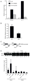
Notch 1 regulates RORγt promoter activity. (A) In-vitro ILCHO treatment down-regulates RORγt and IL-17 mRNA expression. Total RNA was isolated from CD4+ T cells pretreated with 25μM ILCHO or DMSO as a vehicle control and cultured in Th17 polarizing conditions and analyzed by quantitative real time PCR. (B) Human naïve CD4+ T cells (1×10⁷) were nucleoporated with Notch 1 specific siRNA or scrambled siRNA followed by in-vitro Th17 polarization. Cells were harvested and RORγt expression was determined by quantitative RT-PCR. Transcript abundance was normalized to 18s rRNA expression. (C) Schematic representation of putative CSL binding sites in human RORγt promoter. (D) Specific primers were used to amplify putative CSL binding sites on human RORγt promoter. ChIP assay was performed to determine recruitment of Notch1 on RORγt promoter. Data shown represents fold recruitment of Notch1 on CSL binding sites on human RORγt promoter with respect to control IgG normalized with 1% input DNA. Semiquantitative PCR was also performed using 2μl of DNA eluates using specific primers against CSL sites in RORγt promoter to confirm transcript size. Data represents mean± SD of three independent experiments done in triplicates. * p≤0.05, **p≤0.01.
FIGURE 5
FIGURE 5
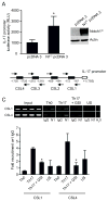
Notch1 regulates human IL-17 promoter activity. (A) 293 T cells were co-transfected with interacellular activated Notch expression vector construct (Notch1IC) along with a human IL-17 promoter construct cloned upstream of firefly luciferase gene. Luciferase assay was performed and data was normalized to renilla luciferase depicted as relative luciferase units (RLU). (B) Schematic representation of putative CSL binding sites in human IL-17 promoter. (C) ChIP assay was performed to determine the recruitment of Notch1 on human IL-17 promoter. Data shown represents fold recruitment of Notch1 on human IL-17 promoter with respect to isotype control IgG normalized to input DNA. Semiquantitative PCR was also performed (2μl of DNA eluates) using specific primers against different putative CSL binding sites in human IL-17 promoter to confirm transcript size. Data shown here represents the mean±SD of three independent experiments done in triplicates.*p≤0.05.
FIGURE 6
FIGURE 6
GSI treatment reduces EAE-induced inflammation and the development of PLP139–151-specific Th17 responses. (A) Clinical scores of SJL/J mice given GSI formulated chow, at 2.5mg/kg alternated with 5mg/kg for 4 weeks. Control mice were given regular chow. n=5 mice in each group. Results represent the mean disease score grouping each group. Splenocytes (B) and cells from the spinal cords (C) of EAE-induced mice were restimulated ex-vivo with PLP139–151 at increasing concentrations and cultured for 5 days. The restimulation supernatants were then analyzed for IL-17 by ELISA. (D) ELISA of IFNγ was performed on PLP139–151 restimulated splenocytes. (E) Total spinal cord cells were stained with CD4 and CD8 antibodies and analyzed by flow cytometry. (F) Intracellular staining of IL-17A in splenocytes of SJL/J mice fed with GSI or control chow. The data shown represent the mean fluorescent intensity of IL-17 in CD4+ T cells. ***p≤0.001

Similar articles

Cited by

References

    1. Weaver CT, Hatton RD, Mangan PR, Harrington LE. IL-17 family cytokines and the expanding diversity of effector T cell lineages. Annu Rev Immunol. 2007;25:821–852. - PubMed
    1. Bettelli E, Korn T, Kuchroo VK. Th17: the third member of the effector T cell trilogy. Curr Opin Immunol. 2007;19:652–657. - PMC - PubMed
    1. Cua DJ, Sherlock J, Chen Y, Murphy CA, Joyce B, Seymour B, Lucian L, To W, Kwan S, Churakova T, Zurawski S, Wiekowski M, Lira SA, Gorman D, Kastelein RA, Sedgwick JD. Interleukin-23 rather than interleukin-12 is the critical cytokine for autoimmune inflammation of the brain. Nature. 2003;421:744–748. - PubMed
    1. Langrish CL, Chen Y, Blumenschein WM, Mattson J, Basham B, Sedgwick JD, McClanahan T, Kastelein RA, Cua DJ. IL-23 drives a pathogenic T cell population that induces autoimmune inflammation. J Exp Med. 2005;201:233–240. - PMC - PubMed
    1. Matusevicius D, Kivisakk P, He B, Kostulas N, Ozenci V, Fredrikson S, Link H. Interleukin-17 mRNA expression in blood and CSF mononuclear cells is augmented in multiple sclerosis. Mult Scler. 1999;5:101–104. - PubMed

Publication types

MeSH terms