. 2011 Jun 15;25(12):1275-88.
doi: 10.1101/gad.2060811.

A Smad action turnover switch operated by WW domain readers of a phosphoserine code

Affiliations

A Smad action turnover switch operated by WW domain readers of a phosphoserine code

Eric Aragón et al. Genes Dev. .

Abstract

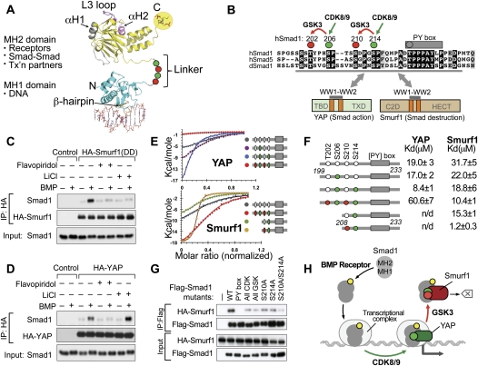
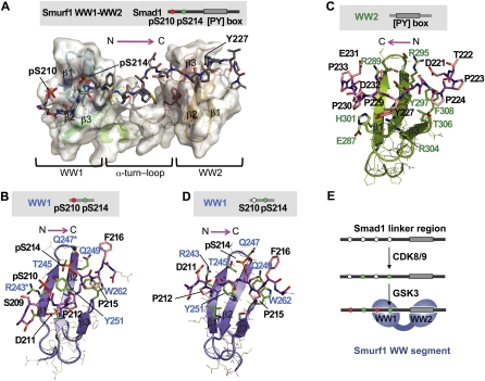
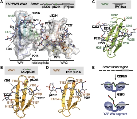
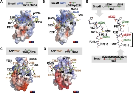
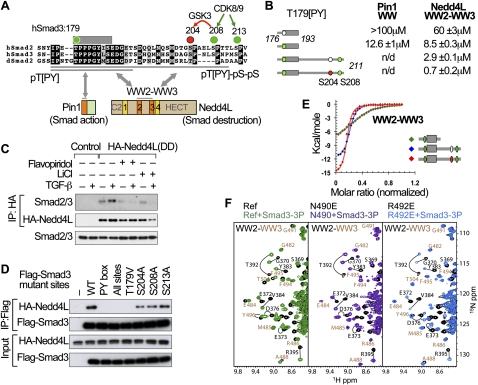
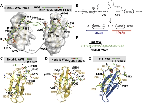
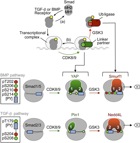
When directed to the nucleus by TGF-β or BMP signals, Smad proteins undergo cyclin-dependent kinase 8/9 (CDK8/9) and glycogen synthase kinase-3 (GSK3) phosphorylations that mediate the binding of YAP and Pin1 for transcriptional action, and of ubiquitin ligases Smurf1 and Nedd4L for Smad destruction. Here we demonstrate that there is an order of events-Smad activation first and destruction later-and that it is controlled by a switch in the recognition of Smad phosphoserines by WW domains in their binding partners. In the BMP pathway, Smad1 phosphorylation by CDK8/9 creates binding sites for the WW domains of YAP, and subsequent phosphorylation by GSK3 switches off YAP binding and adds binding sites for Smurf1 WW domains. Similarly, in the TGF-β pathway, Smad3 phosphorylation by CDK8/9 creates binding sites for Pin1 and GSK3, then adds sites to enhance Nedd4L binding. Thus, a Smad phosphoserine code and a set of WW domain code readers provide an efficient solution to the problem of coupling TGF-β signal delivery to turnover of the Smad signal transducers.


Figures

Figure 1.
Figure 1.
GSK3 switches the binding preference of Smad1 from YAP to Smurf1. (A) Schematic representation of the Smad protein domains and their main functions. The MH1 domain (cyan) contains a β hairpin that mediates binding to dsDNA (orange) (PDB code: 1MHD) (Shi et al. 1998). The MH2 domain (yellow) binds to the type I TGF-β receptor, which involves the L3 loop (magenta); to Smad4 via the phosphorylated C terminus (highlighted) and the α-helix 1 (gray); and to various DNA-binding cofactors and histone-modifying enzymes (PDB code: 1KHX) (Wu et al. 2001). The interdomain linker region (dotted line) contains CDK8/9 and GSK3 phosphorylation sites, represented by green and red circles, respectively. (B) Sequence alignment of the linker region of human Smad1 and Smad5 and Drosophila MAD (dSmad1) proteins, with conserved residues highlighted. The conserved CDK8/9 sites (green) and CDK8/9-primed GSK3 sites (red) and the PY box are shown. The Smad1 (199–232) segment used in this study is underlined. The domain composition of Smurf1 and YAP proteins and the regions that mediate binding to linker phosphorylated Smad1 are indicated. (C) BMP-dependent formation of a complex between HA-Smurf1(DD) and endogenous Smad1 in HEK293 cells, and effects of flavopiridol and LiCl on the formation of this complex. (D) BMP-dependent formation of a complex between HA-YAP and endogenous Smad1 in HEK293 cells, and effects of flavopiridol and LiCl on the formation of this complex. (E) ITC curves for the binding of Smurf1 and YAP WW1–WW2 segments to Smad1 synthetic peptides. (F) Synthetic Smad1 (phospho-)peptides and their affinity for recombinant WW1–WW2 segments of YAP and Smurf1. Colored circles denote phosphorylation of the residues. (G) Effect of alanine mutations in the PY box and the indicated phosphorylation sites on the ability of Flag-tagged Smad1 constructs to bind HA-Smurf1(DD) in HEK293 cells. (H) Schematic summary of the Smad action turnover switch operated by CDK8/9 and GSK3 in combination with YAP and Smurf1.
Figure 2.
Figure 2.
Structure of the Smurf1 WW1–WW2 segment bound to the Smad1 linker. (A) NMR model of the complex between the human Smurf1 WW1–WW2 pair (residues 232–314) and the 208–233 segment of the Smad1 linker diphosphorylated at S210 and S214. Smurf1 is shown as a semitransparent surface, with all elements of secondary structure represented. The Smad1 peptide is shown with a stick representation, with the backbone colored in gray. There are several relative orientations of the WW domains that satisfy all experimental NMR restraints (shown in Supplemental Fig. 2), and, due to this, we call this complex the NMR model. (B) Detailed view of the refined structure of the Smurf1 WW1 domain (slate) bound to the diphosphorylated pS210/pS214 region of the Smad1 linker. Key residues in Smad1 (black) and Smurf1 (blue) are indicated. (Asterisks) Three residues that, when jointly mutated to alanine, decreased the binding affinity of the complex by ∼25-fold. (C) Detailed view of the refined structure of the Smurf1 WW2 domain (green) bound to the PY motif of Smad1. Key residues in Smad1 (black) and Smurf1 (green) are indicated. (D) Detailed view of the refined structure of the Smurf1 WW1 domain (slate) bound to the monophosphorylated pS214 region of the Smad1 linker. (E) Schematic representation of the mode of binding of Smurf1 to the Smad1 linker region.
Figure 3.
Figure 3.
Structure of the YAP WW1–WW2 pair bound to the Smad1 linker. (A) NMR model of the complex between the human YAP WW1–WW2 pair (residues 163–266) and the 199–233 segment of the Smad1 linker diphosphorylated at S206 and S214. YAP is shown as a semitransparent surface, and Smad1 is shown as gray sticks. (B) Detailed view of the refined structure of the YAP WW1 domain (gold) bound to the mono-pS206 phosphorylation site of Smad1 (gray). Key residues in Smad1 (black) and YAP (brown) are indicated. (C) Detailed view of the refined structure of the YAP WW2 domain (green) bound to the PY motif region of Smad1 (gray), with the key residues indicated. (D) Detailed view of the refined structure of the YAP WW1 domain (gold) bound to the diphosphorylated pT202, pS206 region of the Smad1 linker. (E) Schematic representation of the mode of binding of YAP to the Smad1 linker region.
Figure 4.
Figure 4.
The GSK3 phosphorylated Smad1 linker prevents YAP binding. (A,B) Charge distribution on the surface of the Smurf1WW1 domain in complex with the Smad1 linker monophosphorylated at S214 (A) or diphosphorylated at S210 and S214 (B). Negatively charged patches are shown in red, and positively charged patches are shown in dark blue. Smurf1 WW1 is shown as a semitransparent surface, and Smad1 is shown as green sticks. Key residues in Smad1 (black) and Smurf1 WW1 (blue) are shown. The complex is shown in the same orientation as that of Figure 2. (C,D) Charge distribution on the surface of the YAP WW1 domain in complex with the Smad1 linker monophosphorylated at S206 (C) or diphosphorylated at T202 and S206 (D). The YAP WW1 domain is shown as a semitransparent surface and with the same orientation as in Figure 3. The position of T202 is shown in a box. The conformational change observed in pT202 is represented with an arrow. (E) Molecular simulations performed on two peptides corresponding to Smad1 phosphorylated at S206 and S214 (left) or at T202, S206, S210, and S214 (right). Key residues are labeled.
Figure 5.
Figure 5.
Elements of a Smad action turnover switch in the TGF-β pathway. (A) Sequence alignment of the linker regions of human Smad2 and Smad3 and Drosophila Smad2, with the conserved residues highlighted. The conserved CDK8/9 sites (green) and CDK8/9-primed GSK3 site (red) and the PY box are shown. Two Smad3 segments (176–193 and 176–211) used in this study are underlined. The domain composition of Pin1 and Nedd4L proteins and the regions that mediate binding to linker phosphorylated Smad3 are indicated. (B) Synthetic Smad3 (phospho-)peptides and their affinity for the recombinant Pin1 WW domain and Nedd4L WW2–WW3 pair. Colored circles denote phosphorylation of the indicated residues. (n.d.) Not determined. (C) TGF-β-dependent formation of a complex between HA-Nedd4L(DD) and endogenous Smad3, and effects of flavopiridol and LiCl on the formation of this complex. (D) Effect of alanine mutations in the PY box and the indicated phosphorylation sites on the ability of Flag-tagged Smad3 constructs to bind HA-Nedd4L(DD) in HEK293 cells. (E) ITC curves and corresponding fitting to pairs of Nedd4L WW domains and the indicated Smad3 (phospho-)peptides. (F) NMR titrations of WW2–WW3 pairs (wild type in green) with point mutations introduced in two residues that coordinate the pS204pS208 site (violet and royal blue). Residues that belong to WW2 and WW3 are labeled in black and camel, respectively.
Figure 6.
Figure 6.
Basis for Nedd4L and Pin1 recognition of the phosphorylated Smad3 linker. (A) Model of the complex structure between the human Nedd4L WW2–WW3 pair (residues 364–512) and the 176–211 segment of the Smad3 linker triphosphorylated at T179, S204, and S208. Nedd4L is shown as a semitransparent surface, and Smad3 is shown as green sticks. Smad3 residues involved in the interaction with the Nedd4L WW2–WW3 pair are indicated. The 80-amino-acid region connecting the WW2 and WW3 domains (dotted line) does not adopt a defined secondary or tertiary structure, as indicated by near-random 13C chemical shifts. Due to the complexity that this long unstructured part adds to the calculation of the complex, the model has been calculated using three independent molecules (WW2, WW3, and the Smad3 peptide) without the 80-amino-acid region. Three possible orientations of the WW2 and WW3 pair were obtained using a set of RDC experiments. The orientation that yields the best view of the bound Smad3 peptide is shown here. In the bound Smad3, the segment between S186 and G203 does not contact either WW2 or WW3 and is not represented. (B) Schematic of the protein ligation strategy employed to prepare the WW2–WW3 module as two separate fragments for sequential isotope labeling. A fully deuterated WW2 and connector (WW2-conn) segment of Nedd4L was ligated to the protonated WW3 domain, as shown. Using this strategy, the signals from the first part of the protein were filtered, and the analysis of data corresponding to the WW3 domain bound to Smad3 and of that of Smad3 itself was simplified. In the calculation of the complex, this information was combined with that of the WW2 in complex with the pT[PY] peptide and with residual dipolar coupling data obtained from the entire WW2–WW3 segment. (C) Detailed view of the Nedd4L WW2 domain (gold) bound to the phosphorylated PY motif (pT179[PY]) of Smad3 (green). Key residues in Smad3 (black) and Nedd4L (brown) are indicated. This complex has been refined using the data of the WW2–WW3 pair that corresponds to the second WW and the fragment of 176–190 of Smad3. (Asterisks) Two residues that, when jointly mutated to alanine, decreased the binding affinity of the complex by ∼11-fold. (D) Detailed view of the Nedd4L WW3 domain (gold) bound to the diphosphorylated pS204–pS208 sites of Smad3 (green). Key residues in Smad3 (black) and Nedd4L (brown) are indicated. This complex has been refined using the data of the WW2–WW3 that corresponds to the WW3 site and the fragment of 203–211 of Smad3. (E) Solution structure of the Pin1 WW domain bound to the Smad3 pT179[PY] motif. The WW domain is shown as a ribbon representation, shown in marine. Key residues in Smad3 (black) and Pin1 (light brown) are indicated. This complex is displayed using the same orientation as that of the Nedd4L WW2 complex (shown in C) to highlight that these WW domains bind to the pT179[PY] site in opposite orientations. (F) Representation of the distinct portions of the pT179[PY] motif of Smad3 that provide contacts with Pin1 and Nedd4L.
Figure 7.
Figure 7.
The Smad action turnover switch in the BMP and TGF-β pathways: pSer codes and WW domain code readers. (Top panel) Schematic summary of the Smad action turnover switch in the BMP and TGF-β pathways. Following receptor-mediated phosphorylation (yellow circle), Smad proteins translocate to the nucleus and assemble transcriptional complexes, which are phosphorylated at CDK8/9 sites (green circle) in the MH1–MH2 interdomain linker region. This phosphorylation creates high-affinity binding sites for transcriptional partners (such as YAP in the case of the BMP mediator Smad1, Pin1 in the case of the TGF-β mediator Smad3, and probably others), thus achieving peak transcriptional action. Phosphorylation by CDK8/9 also primes the Smads for GSK3-mediated phosphorylation (red symbol) at the −4 position, which favors the binding of ubiquitin ligases Smurf1 (BMP pathway) and Nedd4L (TGF-β pathway), leading to proteasome-dependent degradation of Smad molecules that participate in transcription (erase symbol). Alternatively, C-terminal Smad phosphatases (a) and linker phosphatases (b) reverse these phosphorylation states. See the text for details and citations. (Bottom panels) Schematic of the Smad linker phospho-amino acid codes (insets) and WW domain code readers. The conserved CDK8/9 phosphorylation sites (green circles) and GSK3 sites (red circles) are located at the indicated positions relative to the PY box (slate box). Amino acid positions correspond to Smad1 and Smad3. In the BMP pathway, the YAP WW1 domain binds to pS206 in Smad1, as long as p210 is not phosphorylated. The Smurf1 WW1 domain binds with higher affinity to the pS210–pS214 motif. The WW2 domains bind the [PY] motif. In the TGF-β pathway, the sole WW domain of Pin1 binds the pT179[PY] motif, as does the WW2 domain of Nedd4L. However, the Nedd4L WW3 domain increases the binding affinity by recognizing the pS204–pS208 motif. See the text for additional details and citations on the known roles of these WW domain proteins in Smad signal transduction.

References

    1. Alarcon C, Zaromytidou AI, Xi Q, Gao S, Yu J, Fujisawa S, Barlas A, Miller AN, Manova-Todorova K, Macias MJ, et al. 2009. Nuclear CDKs drive Smad transcriptional activation and turnover in BMP and TGF-β pathways. Cell 139: 757–769 - PMC - PubMed
    1. Baca M, Muir TW, Schnölzer M, Kent SBH 1995. Chemical ligation of cysteine- containing peptides: synthesis of a 22 kDa tethered dimer of HIV-1 protease. J Am Chem Soc 116: 10797–10798
    1. Bartels C, Xia T-H, Billeter M, Güntert P, Wüthrich K 1995. The program XEASY for computer-supported NMR spectral analysis of biological macromolecules. J Biol NMR 5: 1–10 - PubMed
    1. Batut J, Schmierer B, Cao J, Raftery LA, Hill CS, Howell M 2008. Two highly related regulatory subunits of PP2A exert opposite effects on TGF-β/Activin/Nodal signalling. Development 135: 2927–2937 - PMC - PubMed
    1. Brünger AT, Adams PD, Clore GM, DeLano WL, Gros P, Grosse-Kunstleve RW, Jiang JS, Kuszewski J, Nilges M, Pannu NS, et al. 1998. Crystallography and NMR system: a new software suite for macromolecular structure determination. Acta Crystallogr D Biol Crystallogr 54: 905–921 - PubMed

Publication types

MeSH terms

LinkOut - more resources