. 2011 Jun 15;25(12):1320-7.
doi: 10.1101/gad.2053211.

Analysis of protein dynamics at active, stalled, and collapsed replication forks

Affiliations

Analysis of protein dynamics at active, stalled, and collapsed replication forks

Bianca M Sirbu et al. Genes Dev. .

Abstract

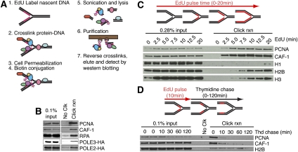
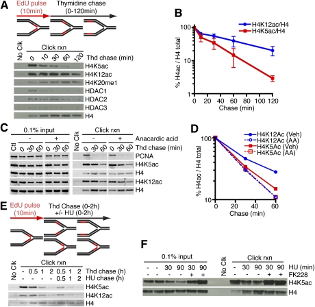
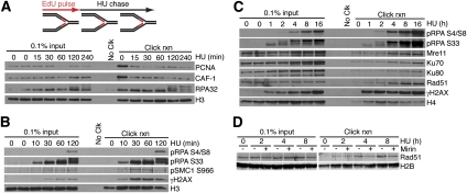
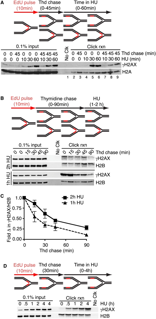
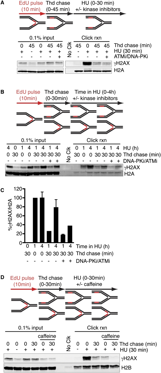
Successful DNA replication and packaging of newly synthesized DNA into chromatin are essential to maintain genome integrity. Defects in the DNA template challenge genetic and epigenetic inheritance. Unfortunately, tracking DNA damage responses (DDRs), histone deposition, and chromatin maturation at replication forks is difficult in mammalian cells. Here we describe a technology called iPOND (isolation of proteins on nascent DNA) to analyze proteins at active and damaged replication forks at high resolution. Using this methodology, we define the timing of histone deposition and chromatin maturation. Class 1 histone deacetylases are enriched at replisomes and remove predeposition marks on histone H4. Chromatin maturation continues even when decoupled from replisome movement. Furthermore, fork stalling causes changes in the recruitment and phosphorylation of proteins at the damaged fork. Checkpoint kinases catalyze H2AX phosphorylation, which spreads from the stalled fork to include a large chromatin domain even prior to fork collapse and double-strand break formation. Finally, we demonstrate a switch in the DDR at persistently stalled forks that includes MRE11-dependent RAD51 assembly. These data reveal a dynamic recruitment of proteins and post-translational modifications at damaged forks and surrounding chromatin. Furthermore, our studies establish iPOND as a useful methodology to study DNA replication and chromatin maturation.


Figures

Figure 1.
Figure 1.
Development of the iPOND technology. (A) iPOND begins by adding EdU to cultured cells. The cells are then treated with formaldehyde to cross-link protein–DNA complexes, washed, and permeabilized with detergent. Copper catalyzes the cycloaddition of biotin-azide to the EdU-labeled DNA. The cells are then lysed in denaturing conditions with sonication. The biotin-labeled DNA–protein complexes are purified using streptavidin-coated beads, cross-links are reversed, and the eluted proteins are analyzed by immunoblotting or other methods like mass spectrometry. (B) Cells were incubated with EdU for 10 min prior to performing iPOND. Cells expressing POLE2-HA or POLE3-HA were used to detect these proteins with the HA antibody. (C) Cells were incubated in EdU-containing medium for increasing times prior to performing the iPOND protocol. (D) Cells were incubated with EdU for 10 min. The EdU-containing medium was removed and cells were washed once before incubating for increasing times in medium containing 10 μM thymidine prior to performing iPOND. In all experiments, the No Clk control is the input sample in the first lane processed with no biotin-azide.
Figure 2.
Figure 2.
HDACs are enriched at replication forks and deacetylate newly deposited histone H4 regardless of fork movement. (A–E). Cells were labeled with EdU for 10 min followed, by a chase into thymidine-containing medium for the indicated times prior to performing iPOND. (B) Quantitation of H4 acetylation levels compared with total H4 in the click reaction samples from three independent experiments. Error bars in all figures are standard deviations. (C,D) Anacardic acid (30 μM) was added to the indicated samples. (E) HU (3 mM) was added to the indicated samples. (F) Cells labeled with EdU were chased into 3 mM HU medium with or without 100 nM FK228 prior to performing iPOND.
Figure 3.
Figure 3.
iPOND monitors post-translational modifications and recruitment of DDR proteins to stalled and collapsed replication forks. (A–D) Cells were labeled with EdU for 15 min (A) or 10 min (B–D), followed by a chase into HU for the indicated times prior to performing iPOND. (D) HU-treated cells were additionally coincubated with or without the Mre11 inhibitor mirin (100 μM) as indicated.
Figure 4.
Figure 4.
γH2AX spreads from a stalled replication fork. (A–D) Cells labeled with EdU for 10 min were chased into thymidine-containing medium prior to addition of HU, then processed using iPOND. The length of the thymidine and HU treatments is indicated. Quantitation of the click reaction samples in C at the 2-h HU-treated samples is from three independent experiments, and at the 1-h HU-treated samples is from two independent experiments.
Figure 5.
Figure 5.
Checkpoint kinases propagate H2AX phosphorylation from stalled replication forks. (A–C) Cells labeled with EdU for 10 min were chased into thymidine, followed by treatment with HU. The length of thymidine and HU treatments are indicated. DNA-PK (KU7441, 1 μM) and ATM (KU5593, 10 μM) inhibitors were added at the same time as HU in the indicated samples. (C) Quantitation of the click reaction samples is the average from two independent experiments and is normalized to the 1-h HU treatment. (D) Cells labeled with EdU for 10 min were chased into thymidine for either 0 or 30 min, followed by a 30-min treatment with HU. Caffeine (10 mM) was added at the same time as HU in the indicated samples.

References

    1. Ahmad A, Takami Y, Nakayama T 1999. WD repeats of the p48 subunit of chicken chromatin assembly factor-1 required for in vitro interaction with chicken histone deacetylase-2. J Biol Chem 274: 16646–16653 - PubMed
    1. Anantha RW, Vassin VM, Borowiec JA 2007. Sequential and synergistic modification of human RPA stimulates chromosomal DNA repair. J Biol Chem 282: 35910–35923 - PubMed
    1. Berkovich E, Monnat RJ Jr, Kastan MB 2007. Roles of ATM and NBS1 in chromatin structure modulation and DNA double-strand break repair. Nat Cell Biol 9: 683–690 - PubMed
    1. Berkovich E, Monnat RJ Jr, Kastan MB 2008. Assessment of protein dynamics and DNA repair following generation of DNA double-strand breaks at defined genomic sites. Nat Protoc 3: 915–922 - PubMed
    1. Bhaskara S, Knutson SK, Jiang G, Chandrasekharan MB, Wilson AJ, Zheng S, Yenamandra A, Locke K, Yuan JL, Bonine-Summers AR, et al. 2010. Hdac3 is essential for the maintenance of chromatin structure and genome stability. Cancer Cell 18: 436–447 - PMC - PubMed

Publication types