. 2011 Sep 22;118(12):3426-35.
doi: 10.1182/blood-2010-11-320812. Epub 2011 Jun 17.

G-CSF supplementation with chemotherapy can promote revascularization and subsequent tumor regrowth: prevention by a CXCR4 antagonist

Affiliations

G-CSF supplementation with chemotherapy can promote revascularization and subsequent tumor regrowth: prevention by a CXCR4 antagonist

Tali Voloshin et al. Blood. .

Abstract

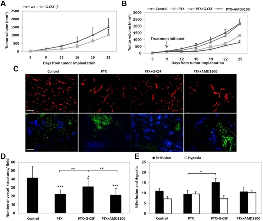
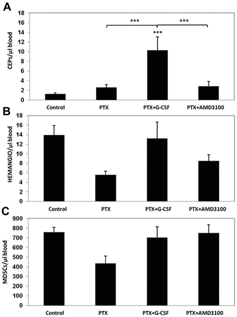
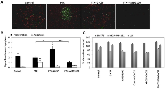
Recombinant granulocyte colony-stimulating factor (G-CSF) is used to accelerate recovery from chemotherapy-induced myelosuppression. G-CSF has been recently shown to stimulate angiogenesis mediated by several types of bone marrow-derived cell populations. To investigate whether G-CSF may alter tumor response to therapy, we studied Lewis lung and EMT/6 breast carcinomas in mice treated with paclitaxel (PTX) chemotherapy in combination with G-CSF. We compared the results obtained to mice treated with PTX and AMD3100, a small-molecule drug antagonist of CXCR4 which, like G-CSF, can be used to mobilize hematopoietic cells. We show that PTX combined with G-CSF treatment facilitates revascularization, leading to an improvement in blood perfusion in LLC tumors, and a decrease in hypoxia in EMT/6 tumors, thus enhancing tumor growth in comparison to PTX or PTX and AMD3100 therapies. We found that hemangiocytes but not Gr-1(+) CD11b(+) cells colonize EMT/6 tumors after treatment with PTX and G-CSF, but not PTX and AMD3100, and therefore may contribute to angiogenesis. However, increases in hemangiocyte colonization were not observed in LLC PTX and G-CSF-treated tumors, suggesting distinct mechanisms of tumor revascularization after G-CSF. Overall, our observations suggest that despite its known considerable clinical benefits, G-CSF might contribute to tumor revascularization by various mechanisms, and diminish the antitumor activity of chemotherapy, an effect that can be prevented by AMD3100.


Figures

Figure 1
Figure 1
LLC tumor growth in G-CSF−/− or G-CSF wt mice treated with PTX, PTX and G-CSF, or PTX and AMD3100 and the relative microvessel density, hypoxia, and perfusion. LLC cells in the quantity of 5 × 10⁵ cells were implanted in the flanks of (A) 8- to 10-week-old G-CSF+/+ or G-CSF−/− 129Sv/C57Bl/6 mice (n = 4-5 mice per group) or (B) 8- to 10-week-old C57Bl/6 which were treated with PTX, PTX and G-CSF, PTX and AMD3100. When tumors reached 200 mm3, treatment was initiated. Tumors were measured regularly using Vernier calipers, and tumor growth (volume) was plotted against number of days after tumor cell implantation. In a parallel experiment, C57Bl/6 mice bearing 500 mm3 LLC tumors (n = 4-5 mice per group) were treated with PTX or PTX in combination with either G-CSF or AMD3100. Tumors were removed 3 days later and evaluated for (C) microvessel density using CD31 immunostaining as a marker for endothelial cells (red; scale bar = 100 μm, 20×/0.50 NA); and hypoxia (green) and vessel perfusion (blue) using hypoxic probe and Hoechst as described in “Quantitation and visualization of tissue hypoxia, vessel perfusion, microcessel density, tumor cell proliferation, and apoptosis” (scale bars = 200 μm; 10×/0.30 NA). Quantification of (D) microvessel density, and (E) perfusion and hypoxia is provided.
Figure 2
Figure 2
Evaluation of BMDC circulating populations after treatment with PTX or PTX in combination with either G-CSF or AMD3100. Non–tumor-bearing C57Bl/6 mice (n = 4 to 5 mice per group) were treated with PTX, PTX and G-CSF, or PTX and AMD3100. Mice were bled via retro-orbital sinus for the evaluation of (A) viable CEPs; (B) hemangiocytes; and (C) Gr-1+/CD11b+ cells (MDSCs), using flow cytometry. ***P < .001 from control group, unless indicated otherwise.
Figure 3
Figure 3
Homing and colonization of BMDCs in LLC and EMT/6 tumors after treatment with PTX or PTX in combination with either G-CSF or AMD3100. (A) C57Bl/6 mice bearing 500 mm3 LLC tumors (n = 4-5 mice per group) or (B) BALB/c mice bearing 500 mm3 EMT/6 murine breast carcinoma (n = 4-5 mice per group) were treated with PTX or PTX in combination with either G-CSF or AMD3100. Tumors were removed 3 days later and prepared as single cell suspensions for the evaluation of hemangiocytes (HEMANGIO) and MDSCs colonization of tumors using flow cytometry. *.05 > P > .01; **.01 > P > .001 from control group, unless indicated otherwise.
Figure 4
Figure 4
Relative SDF-1α protein expression and Bv8 mRNA expression in LLC tumors after treatment with PTX or PTX in combination with either G-CSF or AMD3100. (A) C57Bl/6 mice bearing 500 mm3 LLC tumors (n = 4-5 mice per group) were treated with PTX or PTX in combination with either G-CSF or AMD3100. Tumors were removed 3 days later and sections evaluated for SDF-1α expression (green; scale bar = 500 μm). (B) Quantification of relative SDF-1α expression is provided. (C) In parallel, mRNA was extracted from the same tumors, and Bv8 mRNA levels were measured by qRT-PCR. ***P < .001 from control group, unless indicated otherwise.
Figure 5
Figure 5
Evaluation of tumor cell proliferation and apoptosis in LLC tumors after treatment with PTX or PTX in combination with either G-CSF or AMD3100. (A) C57Bl/6 mice bearing 500 mm3 LLC tumors (n = 4-5 mice per group) were treated with PTX or PTX in combination with either G-CSF or AMD3100. Tumors were removed 3 days later and evaluated for proliferation (red) and apoptosis (green; scale bar = 200 μm). (B) Quantification of perfusion and hypoxia is expressed as percentage. (C) EMT/6, MDA-MB-231, and LLC cells were cultured in the presence of G-CSF or AMD3100 with or without CoCl2. Cell proliferation was evaluated quantitatively with the metabolic indicator dye alamarBlue. Results are presented as the percentage of alamarBlue reduction and were corrected to background values of negative controls.
Figure 6
Figure 6
Analysis of VEGF-A, and MMP-2 levels in tumors after treatment with PTX, PTX and G-CSF or PTX and AMD3100. (A,C) C57Bl/6 mice bearing 500 mm3 LLC tumors (n = 3 mice per group) or (B,D) BALB/c mice bearing 500 mm3 EMT/6 tumors (n = 3 mice per group) were treated with PTX or PTX in combination with either G-CSF or AMD3100. Tumors were removed 3 days later and tumor lysates were evaluated for (A-B) MMP-2 expression by zymography, and (C-D) VEGF-A expression by ELISA. Summary of data are presented in graphs.

References

    1. Kerbel RS. Tumor angiogenesis. N Engl J Med. 2008;358(19):2039–2049. - PMC - PubMed
    1. Hudis CA. Trastuzumab–mechanism of action and use in clinical practice. N Engl J Med. 2007;357(1):39–51. - PubMed
    1. Kim JJ, Tannock IF. Repopulation of cancer cells during therapy: an important cause of treatment failure. Nat Rev Cancer. 2005;5(7):516–525. - PubMed
    1. Friberg LE, Henningsson A, Maas H, Nguyen L, Karlsson MO. Model of chemotherapy-induced myelosuppression with parameter consistency across drugs. J Clin Oncol. 2002;20(24):4713–4721. - PubMed
    1. Smith TJ, Khatcheressian J, Lyman GH, et al. 2006 update of recommendations for the use of white blood cell growth factors: an evidence-based clinical practice guideline. J Clin Oncol. 2006;24(19):3187–3205. - PubMed

Publication types

MeSH terms