. 2011 Jun 19;476(7360):336-40.
doi: 10.1038/nature10230.

A forty-kilodalton protein of the inner membrane is the mitochondrial calcium uniporter

Affiliations

A forty-kilodalton protein of the inner membrane is the mitochondrial calcium uniporter

Diego De Stefani et al. Nature. .

Abstract

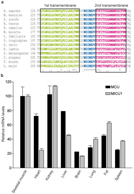
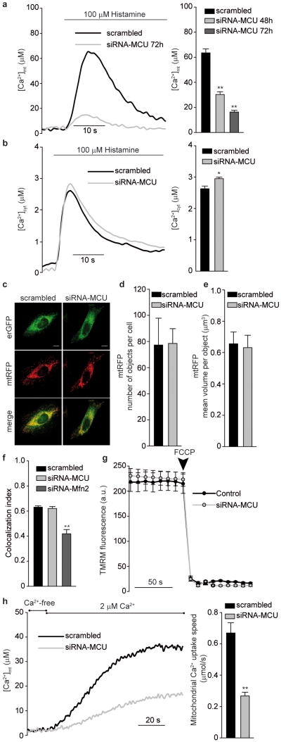
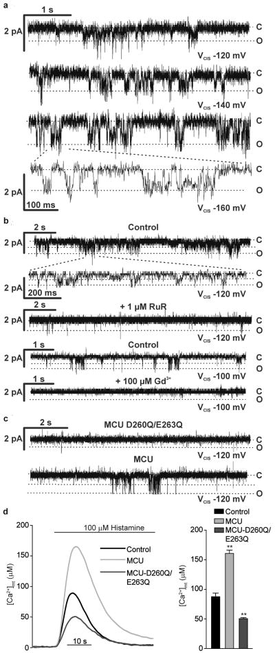
Mitochondrial Ca(2+) homeostasis has a key role in the regulation of aerobic metabolism and cell survival, but the molecular identity of the Ca(2+) channel, the mitochondrial calcium uniporter, is still unknown. Here we have identified in silico a protein (named MCU) that shares tissue distribution with MICU1 (also known as CBARA1), a recently characterized uniporter regulator, is present in organisms in which mitochondrial Ca(2+) uptake was demonstrated and whose sequence includes two transmembrane domains. Short interfering RNA (siRNA) silencing of MCU in HeLa cells markedly reduced mitochondrial Ca(2+) uptake. MCU overexpression doubled the matrix Ca(2+) concentration increase evoked by inositol 1,4,5-trisphosphate-generating agonists, thus significantly buffering the cytosolic elevation. The purified MCU protein showed channel activity in planar lipid bilayers, with electrophysiological properties and inhibitor sensitivity of the uniporter. A mutant MCU, in which two negatively charged residues of the putative pore-forming region were replaced, had no channel activity and reduced agonist-dependent matrix Ca(2+) concentration transients when overexpressed in HeLa cells. Overall, these data demonstrate that the 40-kDa protein identified is the channel responsible for ruthenium-red-sensitive mitochondrial Ca(2+) uptake, thus providing a molecular basis for this process of utmost physiological and pathological relevance.


Conflict of interest statement

Author Information. The authors declare no conflict of interest.

Figures

Figure 1
Figure 1. MCU includes two highly conserved transmembrane domains and is ubiquitously expressed in mammals, similarly to its putative regulator MICU1
a, Alignment of the putative transmembrane domain and pore region of MCU proteins from 14 different species. b, Quantitative Real Time PCR analysis of mouse tissues. mRNA extraction and quantitative PCR as described in the methods section. Expression levels are normalized to skeletal muscle, and presented as means ± S.D. (n = 3).
Figure 2
Figure 2. MCU silencing strongly inhibits mitochondrial Ca2+ uptake without causing morphological rearrangement or changes in the electrochemical gradient
a, [Ca2+]mt and b, [Ca2+]cyt measurements in MCU-silenced cells. c, Fluorescence images of mtRFP- and erGFP-labeled mitochondria and ER, respectively. d and e, Mitochondrial number and volume, as deduced by calculating object number (d) and size (e). f, ER/mitochondria colocalization, estimated by Pearson's correlation coefficient. g, TMRM fluorescence measurements. h, [Ca2+]mt measurements in permeabilized cells. In this and following figures, experiments are representative of >5 trials, conditions are in the methods section, and statistics in Supplementary Table 2. * p<0.05, ** p<0.001
Figure 3
Figure 3. MCU overexpression increases mitochondrial Ca2+ accumulation in intact and permeabilized cells, buffers cytosolic [Ca2+] rises, and sensitizes to apoptotic simuli; GFP-tagged MCU demonstrates mitochondrial localization and suggests a putative membrane topology
a, [Ca2+]mt and b, [Ca2+]cyt measurements in MCU-overexpressing cells. c, [Ca2+]mt measurements in permeabilized cells. d, Cell viability upon apoptotic challenge. e, Subcellular fractionation of MCU-GFP- and mtGFP-expressing cells and Western blotting for GFP, matrix (HSP60) and OMM (VDAC1) markers. f, GFP proteinase K degradation and fluorescence quenching by Trypan blue in permeabilized cells. g, Schematic representation of the predicted MCU topology. RuR, Ruthenium Red. ** p<0.001.
Figure 4
Figure 4. The purified MCU protein exhibits channel activity in lipid bilayers, with the properties of the uniporter; MCUD260Q,E263Q shows no channel activity and reduces [Ca2+]mt transients in cells
a-c Electrophysiological recordings of purified MCU or MCUD260Q,E263Q, produced in E. coli (a and b) or in vitro (c) and reconstituted in planar lipid bilayers. a, MCU traces at different applied voltages; b, MCU traces before and after addition of RuR or Gd3+; c, MCUD260Q,E263Q traces. After 10 minutes recording with no current, MCU was added and channel activity was detected after 1-2 minutes (lower trace). d, [Ca2+]mt measurements in MCUD260Q,E263Q-expressing cells.

Comment in

  • Calcium: mitochondria channel calcium.
    Wrighton KH. Wrighton KH. Nat Rev Mol Cell Biol. 2011 Jul 13;12(8):463. doi: 10.1038/nrm3156. Nat Rev Mol Cell Biol. 2011. PMID: 21750571 No abstract available.

References

    1. Szabadkai G, Duchen MR. Mitochondria: the hub of cellular Ca2+ signaling. Physiology (Bethesda) 2008;23:84–94. - PubMed
    1. Carafoli E. Historical review: mitochondria and calcium: ups and downs of an unusual relationship. Trends Biochem Sci. 2003;28:175–181. - PubMed
    1. Perocchi F, et al. MICU1 encodes a mitochondrial EF hand protein required for Ca(2+) uptake. Nature. 2010;467:291–296. - PMC - PubMed
    1. Berridge MJ, Bootman MD, Roderick HL. Calcium signalling: dynamics, homeostasis and remodelling. Nat Rev Mol Cell Biol. 2003;4:517–529. - PubMed
    1. Rizzuto R, Brini M, Murgia M, Pozzan T. Microdomains with high Ca2+ close to IP3-sensitive channels that are sensed by neighboring mitochondria. Science. 1993;262:744–747. - PubMed

Publication types

MeSH terms