. 2011 Jun 19;17(7):845-53.
doi: 10.1038/nm.2370.

Loss of JAK2 regulation via a heterodimeric VHL-SOCS1 E3 ubiquitin ligase underlies Chuvash polycythemia

Affiliations

Loss of JAK2 regulation via a heterodimeric VHL-SOCS1 E3 ubiquitin ligase underlies Chuvash polycythemia

Ryan C Russell et al. Nat Med. .

Abstract

Chuvash polycythemia is a rare congenital form of polycythemia caused by homozygous R200W and H191D mutations in the VHL (von Hippel-Lindau) gene, whose gene product is the principal negative regulator of hypoxia-inducible factor. However, the molecular mechanisms underlying some of the hallmark abnormalities of Chuvash polycythemia, such as hypersensitivity to erythropoietin, are unclear. Here we show that VHL directly binds suppressor of cytokine signaling 1 (SOCS1) to form a heterodimeric E3 ligase that targets phosphorylated JAK2 (pJAK2) for ubiquitin-mediated destruction. In contrast, Chuvash polycythemia-associated VHL mutants have altered affinity for SOCS1 and do not engage with and degrade pJAK2. Systemic administration of a highly selective JAK2 inhibitor, TG101209, reversed the disease phenotype in Vhl(R200W/R200W) knock-in mice, an experimental model that recapitulates human Chuvash polycythemia. These results show that VHL is a SOCS1-cooperative negative regulator of JAK2 and provide biochemical and preclinical support for JAK2-targeted therapy in individuals with Chuvash polycythemia.


Conflict of interest statement

COMPETING FINANCIAL INTERESTS

The authors declare no competing financial interests.

Figures

Figure 1
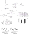
Figure 1. CP-VHL exhibits altered binding to ECV components and JAK2
(A) HEK293 cells transfected with the indicated plasmids were immunoprecipitated and immunoblotted with the indicated antibodies. (B) 35S-radiolabelied 786-O subclones stably expressing indicated HA-VHL were immunoprecipitated with anti-HA antibody, resolved by SDS-PAGE and visualized by autoradiography. (C) HEK293 cells transfected with the indicated combination of plasmids were treated with (+) or without (−) MG132. Equal amounts of cell lysates were immunoprecipitated and immunoblotted with the indicated antibodies. (D) HEK293 cells transfected with the indicated plasmids were lysed in the absence of MG132, immunoprecipitated and immunoblotted with the indicated antibodies. WCE: whole cell extract; IP: immunoprecipitation; IB: immunoblot; AR: autoradiography; Asterisk: non-specific protein bands.
Figure 2
Figure 2. VHL promotes ubiquitin-mediated destruction of pJAK2
(A) HEK293 cells transfected with the indicated plasmids were lysed and immunoblotted with the indicated antibodies. (B) BaF3-EPOR cells infected with retrovirus encoding HA-VHL were lysed, immunoprecipitated with anti-HA or isotype-matched control antibody and immunoblotted with anti-VHL antibody (right panel). The same HA-VHL-expressing BaF3-EPOR cells treated with (+) or without (−) EPO were immunoprecipitated and immunoblotted with the indicated antibodies (left panel). (C) HEK293 cells transfected with the indicated plasmids were treated with (+) or without (−) 20U ml−1 of EPO and pJAK2 isolated via anti-T7 immunoprecipitation (left panel), which was then added to an in vitro ubiquitylation reaction containing proteasome-depleted S100 fractions with (+) or without (−) VHL (right panels). Reaction mixtures were re- immunoprecipitated with anti-T7 antibody and immunoblotted with the indicated antibodies. (D) BaF3-EPOR-shVHL or -shScr cells stimulated with EPO for indicated time periods were lysed and immunoblotted with the indicated antibodies. RNA was also extracted and indicated STAT5- responsive mRNA levels were measured by quantitative real-time PCR normalized to GAPDH and EPO-stimulated levels of transcripts in BaF3-EPOR-shScr cells arbitrarily set to 1.0 (graph). Error bars represent standard deviations of the fold-changes over three independent experiments. (E) HEK293 cells transfected with indicated plasmids were lysed and immunoblotted with the indicated antibodies. (F) Expression profiles of known HIF- (left panel) and STAT5- (right panel) responsive genes were generated by analysis of 18 RCC samples in comparison to normal matched tissue control run on the Affymetrix Human Genome U133B Array (GEO GDS507). Median expression is indicated by horizontal bar. Error bars represent the standard deviation in the levels for each sample set. Asterisk denotes non-specific protein bands.
Figure 3
Figure 3. VHL binds to and cooperates with SOCS1 to negatively regulate pJAK2
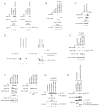
(A,B) HEK293 cells transfected with the indicated plasmids were lysed, immunoprecipitated and immunoblotted with the indicated antibodies. (C) BaF3-EPOR cells stimulated with (+) or without (−) EPO were lysed, immunoprecipitated with anti-VHL or isotype-matched control antibody and immunoblotted with the indicated antibodies. (D) Affinity-purified HA-VHL(C162F) and cell lysates generated from HEK293 cells expressing FLAG-SOCS1 or HA-VHL(C162F) were resolved on SDS-PAGE and immunoblotted with anti-HA antibody (lanes 1–3). HEK293 cells transfected with empty plamid (Mock) or plasmid encoding FLAG-SOCS1 were lysed and immunoblotted with anti-FLAG antibody (lanes 4 and 5) or immunoprecipitated with anti-FLAG antibody and far-Western blotted (FB) with purified recombinant HA-VHL(C162F) followed by anti-HA antibody (lanes 6 and 7). (E) Whole cell extracts prepared from HEK293 cells transfected with the indicated plasmids were immunoblotted with the indicated antibodies. (F) 786-O subclones stably expressing the indicated HA-VHL were lysed and immunoblotted with indicated antibodies (right panel) or immunoprecipitated with anti-HA antibody and immunoblotted with indicated antibodies (left panel). (G) HEK293 cells transfected with the indicated plasmids in combination with a plasmid encoding HA-SOCS1 were lysed, immunoprecipitated and immunoblotted with anti-VHL antibody. (H) Whole cell extracts prepared from HEK293 cells transfected with the indicated plasmids and stimulated with EPO were immunoblotted with the indicated antibodies. Asterisk: non-specific protein bands.
Figure 4
Figure 4. CP-VHL mutants are defective in pJAK2 degradation and pharmacologic JAK2 inhibition attenuates VHL-dependent BaF3-EPOR colony formation
(A) HEK293 cells transfected with the indicated plasmids were lysed, immunoprecipitated and immunoblotted with anti-HA antibody. (B) HEK293 cells transfected with the indicated plasmids were treated with EPO and MG132, lysed, immunoprecipitated and immunoblotted with the indicated antibodies. (C) Equal amounts of T7-pJAK2 isolated from EPO-treated HEK293 cells were mixed with HEK293 cell lysates expressing indicated plasmids. Reaction mixtures were immunoprecipitated with anti-T7 antibody and immunoblotted with anti-pJAK2 antibody. (D) BaF3-EPOR-shVHL cells infected with lentivirus encoding HA-VHL(R200W and WT) were treated with EPO for indicated time periods and immunoblotted with the indicated antibodies. (E) Polyclonal BaF3-EPOR cells stably expressing shVHL or shScr were plated in 1% methylcellulose containing the indicated levels of EPO (top graph) or IL3 (bottom graph). Colony numbers were generated from a single-blind analysis and are representative of four (top graph) or three (bottom graph) independent experiments performed in duplicate. Error bars represent standard deviation between replicate wells. (F) BaF3-EPOR cells were plated in 1% methylcellulose containing EPO in combination with the indicated concentrations of JAK2 inhibitor TG101209. Colony numbers were generated from a single-blind analysis and are representative of two independent experiments performed in triplicate. Error bars represent standard deviation between replicate wells. (G) BaF3-EPOR-shVHL and -shScr cells were plated in 1% methylcellulose containing 100mU EPO in combination with the indicated concentrations of TG101209. Eighty colonies were counted for each condition and average colony size was determined by digitization of microscope photographs followed by measurement of pixel count for each colony, generating an arbitrary two-dimensional area directly proportional to colony size. Scare bar = 0.2mm. Colonies extracted from methylcellulose were lysed and immunoblotted with the indicated antibodies (right panels).
Figure 5
Figure 5. TG101209 treatment rescues polycythemia features in VhlR/R mice
(A) Hct levels of vehicle or TG101209 treated VhlR/R mice. Normal Hct range highlighted in grey. P < 0.05 after 5 weeks of treatment. Error bars = standard error of the mean (SEM). (B) Photographs of the plantar footpads of vehicle and TG101209 treated VhlR/R mice. Scale bar = 3.0mm (C) Photographs of spleens (scale bar = 4.0mm) and photomicrographs of H&E stained sections of spleens (scale bars = 10μm) from VhlR/R mice treated with vehicle or TG10209. Yellow arrows indicate megakaryocytes. (D) Quantification of average number of megakaryocytes per high power field in spleens of vehicle or TG101209 treated VhlR/R mice, P < 0.005. Error bars = SEM. (E) Numbers of CFU-E colonies formed from the splenic haematopoietic precursors of VhlR/R versus WT mice when cultured in the presence of 3U ml−1 of EPO, P < 0.005. Error bars = SEM. (F) Single cell suspensions enriched with erythroid progenitors generated from spleens of phenylhydrazine-treated VhlR/R or WT mice were washed, starved in cytokine-free media and treated with increasing concentrations of exogenous EPO for 15 min. Cell lysates were then immunoblotted with the indicated antibodies. (G) VhlR/R splenic cells were lysed and sonicated in the presence of phosphatase inhibitors and MG132. Lysates were equally divided, immunoprecipitated with anti-VHL or isotype-matched control antibody, and immunoblotted with the indicated antibodies. (H) Numbers of CFU-E colonies formed from the splenic haematopoietic precursors of VhlR/R mice when cultured in the presence of vehicle or TG101209, P < 0.05. Error bars = SEM. Asterisk denotes non-specific protein bands.
Figure 6
Figure 6. The ‘SOCS groove’ and the revised molecular model of CP
(A) Mutations that influence SOCS1 binding are clustered within the ‘SOCS-groove’ as indicated (red) on the VHL/Elongin B/Elongin C crystal structure bound to HIF1α peptide. Analyzed using DeppView/Swiss-PdbViewer v4.0. (B) Molecular model of CP. See text for details.

Similar articles

Cited by

References

    1. Ivan M, et al. HIFalpha targeted for VHL-mediated destruction by proline hydroxylation: implications for O2 sensing. Science. 2001;292:464–8. - PubMed
    1. Jaakkola P, et al. Targeting of HIF-alpha to the von Hippel-Lindau ubiquitylation complex by O2- regulated prolyl hydroxylation. Science. 2001;292:468–72. - PubMed
    1. Maxwell P, et al. The von Hippel-Lindau gene product is necessary for oxgyen-dependent proteolysis of hypoxia-inducible factor α subunits. Nature. 1999;399:271–5. - PubMed
    1. Ohh M, et al. Ubiquitination of hypoxia-inducible factor requires direct binding to the beta-domain of the von Hippel-Lindau protein. Nat Cell Biol. 2000;2:423–7. - PubMed
    1. Roberts AM, Ohh M. Beyond the hypoxia-inducible factor-centric tumour suppressor model of von Hippel-Lindau. Curr Opin Oncol. 2008;20:83–9. - PubMed

Publication types

MeSH terms