. 2011 Jun 19;10(7):545-52.
doi: 10.1038/nmat3049.

Nanoparticles that communicate in vivo to amplify tumour targeting

Affiliations

Nanoparticles that communicate in vivo to amplify tumour targeting

Geoffrey von Maltzahn et al. Nat Mater. .

Abstract

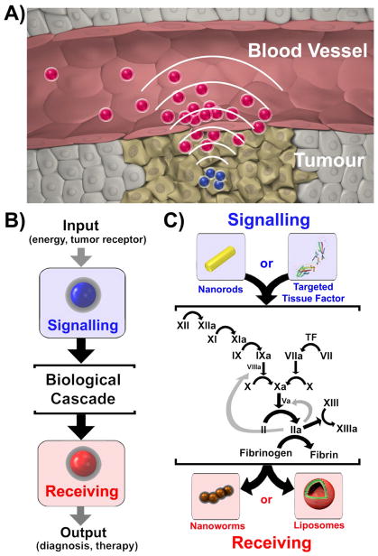
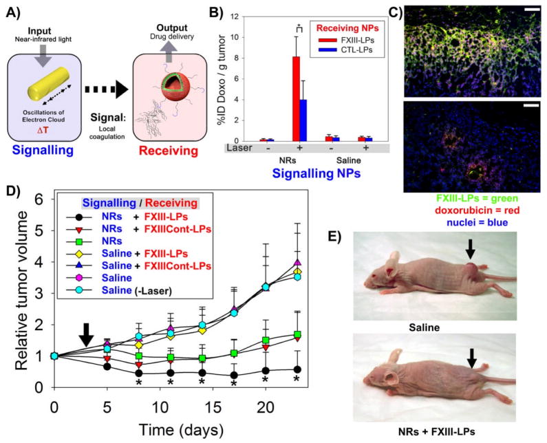
Nanomedicines have enormous potential to improve the precision of cancer therapy, yet our ability to efficiently home these materials to regions of disease in vivo remains very limited. Inspired by the ability of communication to improve targeting in biological systems, such as inflammatory-cell recruitment to sites of disease, we construct systems where synthetic biological and nanotechnological components communicate to amplify disease targeting in vivo. These systems are composed of 'signalling' modules (nanoparticles or engineered proteins) that target tumours and then locally activate the coagulation cascade to broadcast tumour location to clot-targeted 'receiving' nanoparticles in circulation that carry a diagnostic or therapeutic cargo, thereby amplifying their delivery. We show that communicating nanoparticle systems can be composed of multiple types of signalling and receiving modules, can transmit information through multiple molecular pathways in coagulation, can operate autonomously and can target over 40 times higher doses of chemotherapeutics to tumours than non-communicating controls.


Figures

Figure 1
Figure 1. Nanoparticle communication for amplified tumour targeting
A) Schematic representation of communication between system components. Tumour-targeted Signalling nanoparticles broadcast tumour location to Receiving nanoparticles in circulation. B) Harnessing a biological cascade to transmit and amplify nanoparticle communications. C) Molecular signalling pathway between Signalling and Receiving components. Signalling and Receiving components act as unnatural inputs and outputs to the coagulation cascade, respectively. Signalling components are either tumour-targeted plasmonic gold nanorods (NRs), which initiate coagulation cascade activation in tumours by photothermally disrupting tumour vessels and activating the extrinsic and intrinsic coagulation pathways, or tumour-targeted truncated tissue factor proteins, which are latent in circulation and activate the extrinsic coagulation pathway upon binding to tumour receptors. Communication is exploited to recruit inorganic (iron oxide nanoworms) or organic (drug-loaded liposomes) Receiving components via activity of the coagulation transglutaminase FXIII or via targeting of polymerized fibrin.
Figure 2
Figure 2. ‘Signalling’ component characterization
A) Schematic of nanorod-directed coagulation and transmission electron microscopy of near-infrared absorbing nanorods. Gold nanorods (NRs) are targeted to tumours to specify local coagulation cascade activation via photothermal conversion of near-infrared energy. B) Probing the coagulation-dependent and -independent protein tropism to heated tumours. Fibrinogen and albumin were labelled with unique near-infrared fluorochromes and injected into mice bearing bi-lateral MDA-MB-435 tumours. Immediately following injection, one tumour on each mouse was heated using a temperature-controlled water bath. At 24 hrs post-injection, mice were dissected and tumours imaged for the relative abundance of fibrinogen (green) and albumin (red). C). Thermographic imaging of PEG-NR- and saline-injected mice under near-infrared irradiation of the right flank. D) Fluorescence reflectance imaging of mice to visualize fibrinogen tropism to PEG-NR-heated tumours. E) Schematic of tumour-targeted tissue factor stimulation of the coagulation cascade in response to tumour receptors. Signalling components are ligand-targeted, truncated human tissue factor proteins (tTF-RGD) proteins that are latent in circulation and autonomously gain coagulation-inducing activity upon binding to αvβ3 receptors in tumour blood vessels and associating with endothelial cell surface phosphatidylserine. F) Intraoperative images at 24-hrs post-tissue factor injection revealing tTF-RGD-mediated haemorrhaging. G) Probing the coagulation-dependent and -independent protein tropism to tumours on tTF-RGD-injected mice. tTF-RGD Signalling components were injected intravenously at varying doses alongside mixtures of fluorescent fibrinogen (green, VT750) and albumin (red, VT680) to monitor tTF-RGD-mediated coagulation in tumours. H) Histopathologic analysis of tumour fibrinogen distribution without (left) and with 25μg tTF-RGD Signalling component co-injection (right) (Red = CD31 blood vessel stain; Green = injected fibrinogen fluorescence; Blue = nuclear stain; scale bars=100 μm)
Figure 3
Figure 3. ‘Receiving’ component synthesis and testing
A) Schematic of Receiving NP homing to regions of coagulation. Nanoworm (NW) imaging agents and drug-loaded liposomes (LPs) (top and bottom, respectively) were derivatized with coagulation-targeting peptides to form Receiving NPs. B) Nanostructure and targeting ligands of Receiving NPs. Transmission electron microscopy images of the two classes of nanomaterials utilized in Receiving NP synthesis: iron oxide nanoworms (NWs; scale bar=50 nm) and doxorubicin-loaded liposomes (LPs; scale bar=400 nm). Two peptides were utilized to generate Receiving NPs: a fibrin-binding peptide and a glutamine-containing substrate for the coagulation transglutaminase FXIII to respectively direct particle binding and covalent attachment in regions of coagulation. C) Fluorescence reflectance imaging of Receiving NP homing to externally-heated tumours. Mixtures of targeted (green) and untargeted (red) NWs, labelled with the unique NIR-fluorochromes VT750 and VT680, respectively, were intravenously injected into mice bearing bilateral MDA-MB-435 tumours. Immediately following injection, one tumour was submerged in a temperature-controlled water bath for 20 min and mice were dissected at 24 hrs for fluorescent organ imaging. Overlaid fluorescence images are shown for targeted (green) and untargeted (red) Receiving NP accumulation in both heated (+, 45oC heating) and naïve (−) tumours from the same mouse. D) Histopathological analysis of Receiving NP homing to heated tumours. Histological sections from naïve (top) and heated (bottom, 45oC) tumours in FXIII-NW-injected mice were stained for CD31 (red) and nuclei (blue) and imaged to reveal Receiving NP distribution (green). (Scale bars = 100 μm) E) Quantifying the amplification of FXIII-NW and FXIIIControl-NW Receiver homing to heated over unheated tumours. The fold enhancement of NW targeting is plotted across the range of temperatures tested (p=0.02 and 0.03 for the difference between FXIII- -NWs and FXIIIControl-NWs at 45oC and 49oC, respectively; paired, two-sided t-test, n=4; error bars=std. dev.). F) Quantifying the amplification of FXIII-LP and FXIIIControl–LP (Drug-loaded Liposome) Receiver homing to heated over unheated tumours. The fold enhancement of doxorubicin accumulation in tumours is plotted across the range of temperatures tested for FXIII- LPs and FXIIIControl - LPs (p=0.025 and p=0.049 for the difference between FXIII- NWs and FXIIIControl-NWs at 45oC and 49oC, respectively; unpaired, two-sided t-test, n=3; error bars=std. dev.).
Figure 4
Figure 4. Amplified tumour targeting with two systems of communicating nanoparticles
A) Schematic of communicating nanoparticles. B) Experimental timeline for testing communicating nanoparticles. C) Thermographic imaging of photothermal PEG-nanorod heating. At 72 hrs post NR- or saline-injection (10 mg Au/kg), mice were co-injected with coagulation-targeted (FXIII- NWs) and untargeted (FXIIIControl-NWs) and their right flanks were broadly irradiated (810 nm, ~0.75 W/cm2, 20 min) under infrared thermographic surveillance to reveal surface temperatures. D) Overlaid fluorescence reflectance image of targeted and untargeted Receiving NP homing. At 24 hrs post-irradiation, whole-animal fluorescence imaging revealed the distributions of coagulation targeted (FXIII-NWs, green) and untargeted (FXIIIControl-NWs, red) Receiving NPs. E) Quantification of Receiving NP homing in irradiated vs contralateral unirradiated tumours. After whole-animal imaging, mice were dissected and the fluorescence of each tumour was measured to quantify the homing of Receiving NPs. (* indicates p=0.02, paired, two-sided t-test; n=4; error bars=std. dev.) F) Schematic of a nanosystem that communicates autonomously in the presence of tumour receptors. G) Experimental timeline for testing the autonomous nanosystem in vivo. H) Intraoperative imaging of NW Receivers. Nu/nu mice bearing a single MDA-MB-435 tumour were intravenously injected with communicating (tTF-RGD + FXIII-NWs) or control (tTF-RGD + FXIIIControl-NWs) systems, FXIII-NWs alone, or NWs targeted by the peptide used to direct Signalling component tumour homing (1 mg/kg tTF-RGD). At 24 hrs post-injection, tumours were surgically exposed for fluorescent intraoperative imaging of NW homing. (FXIIICont-NWs = FXIIIControl-NWs) I) Tumour specificity of the autonomous nanosystem. Excised organs from mice injected with autonomously communicating nanosystems (tTF-RGD + FXIII-NWs) were imaged for NW fluorescence at 24 hrs post-injection (1 mg/kg tTF-RGD). J) Histopathological analysis of NW Receivers. Histopathological sections from experiments in H). At 24 hrs post-NW injection, mice were sacrificed and tumours were analyzed for NW Receiver distribution in histology. (Red= CD31 blood vessel stain, Blue= DAPI nuclear stain, Green=NW Receiver distribution, RGD-NW scale bar=100 μm; All others = 200 μm)
Figure 5
Figure 5. Amplified tumour therapy with communicating nanoparticles
A) Schematic of therapeutic system of communicating nanoparticles B) Quantification of doxorubicin-loaded LP Receiver homing in irradiated vs contralateral unirradiated tumours. At 96 hrs after Signalling NP injection, mice were dissected and the doxorubicin fluorescence of each tumour homogenate in acidic ethanol was measured to quantify the homing of Receiving NPs. (* indicates p=0.021, unpaired, two-sided t-test, n=4; error bars=std. dev.) C) Histopathological analysis of NR-directed FXIII- LP targeting and doxorubicin delivery. Histopathological sections from the integrated NP signalling experiments in B). At 24 hrs post-NW injection, mice were sacrificed and tumours were analyzed for FXIII-LP and doxorubicin distributions in histology. (Red= doxorubicin, Blue= DAPI nuclear stain, Green=FXIII- LP distribution). (Scale bars = 100 μm). D) Tumour volumes following a single treatment with communicating nanoparticle systems and controls. Tumours in all treatment groups except Saline (laser) were exposed to near-infrared irradiation for 20 min (~0.75 W/cm2, ~810 nm, arrow) 72 hrs after i.v. nanorod or saline injection (p<0.05 for NR + FXIII-LPs and all other treatment sets between days 8 and 24; ANOVA, n=7 mice in each set). E) Representative images of mice treated with communicating nanoparticles (NRs + FXIII-LPs, below) compared with untreated controls (Saline, above) (20 days post-treatment).

Comment in

  • Nanomedicine: swarming towards the target.
    Wang Y, Brown P, Xia Y. Wang Y, et al. Nat Mater. 2011 Jun 19;10(7):482-3. doi: 10.1038/nmat3060. Nat Mater. 2011. PMID: 21685899 No abstract available.
  • Nanotechnology: Tag teams.
    McCarthy N. McCarthy N. Nat Rev Cancer. 2011 Jul 14;11(8):537. doi: 10.1038/nrc3111. Nat Rev Cancer. 2011. PMID: 21753792 No abstract available.

Similar articles

Cited by

References

    1. Chan WC, Nie S. Quantum dot bioconjugates for ultrasensitive nonisotopic detection. Science. 1998;281:2016–2018. - PubMed
    1. Park JH, et al. Magnetic iron oxide nanoworms for tumour targeting and imaging. Advanced Materials. 2008;20:1630. - PMC - PubMed
    1. Xia YN, Halas NJ. Shape-controlled synthesis and surface plasmonic properties of metallic nanostructures. Mrs Bulletin. 2005;30:338–344.
    1. Gref R, et al. Biodegradable long-circulating polymeric nanospheres. Science. 1994;263:1600–1603. - PubMed
    1. Sengupta S, et al. Temporal targeting of tumour cells and neovasculature with a nanoscale delivery system. Nature. 2005;436:568–572. - PubMed

Publication types