. 2011 Jun 19;12(8):770-7.
doi: 10.1038/ni.2050.

Perforin pores in the endosomal membrane trigger the release of endocytosed granzyme B into the cytosol of target cells

Affiliations

Perforin pores in the endosomal membrane trigger the release of endocytosed granzyme B into the cytosol of target cells

Jerome Thiery et al. Nat Immunol. .

Abstract

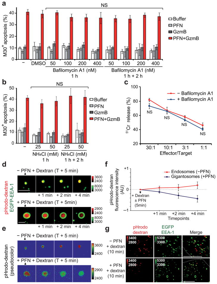
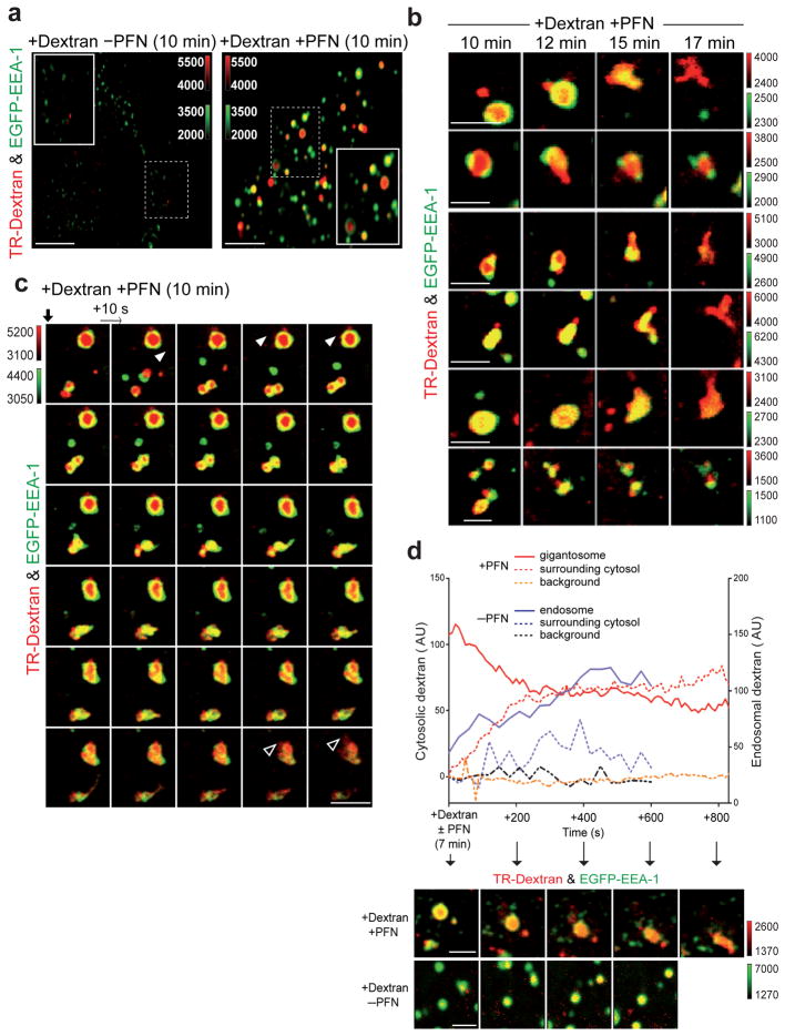
How the pore-forming protein perforin delivers apoptosis-inducing granzymes to the cytosol of target cells is uncertain. Perforin induces a transient Ca2+ flux in the target cell, which triggers a process to repair the damaged cell membrane. As a consequence, both perforin and granzymes are endocytosed into enlarged endosomes called 'gigantosomes'. Here we show that perforin formed pores in the gigantosome membrane, allowing endosomal cargo, including granzymes, to be gradually released. After about 15 min, gigantosomes ruptured, releasing their remaining content. Thus, perforin delivers granzymes by a two-step process that involves first transient pores in the cell membrane that trigger the endocytosis of granzyme and perforin and then pore formation in endosomes to trigger cytosolic release.


Conflict of interest statement

Conflict of interest disclosure

The authors declare no competing financial interests.

Figures

Figure 1
Figure 1. Inhibition of gigantosome formation does not impair GzmB-induced apoptosis
(a–b) HeLa cells transfected with EGFP-Rab5(WT) or EGFP-Rab5(S34N) dominant negative mutant (a, top row) were treated with buffer or sublytic rat PFN ± 100 nM native human GzmB, and apoptosis in EGFP+ cells was measured 2 h later by labeling with M30 mAb (which recognize a cytokeratin-18 epitope, revealed after caspase cleavage). Representative flow cytometry histograms (a) (MFI, mean fluorescence intensity) and mean ± s.d. of percentage of M30+ cells from three independent experiments (b) are shown. P values were determined by unpaired two-tailed student’s t-test. There was no significant (NS) difference in GzmB-mediated apoptosis in Rab5(S34N)-transfected cells relative to Rab5(WT)-transfected cells. (c) Analysis of procaspase-3 activation by immunoblot in HeLa cells transfected with EGFP-Rab5(WT) or EGFP-Rab5(S34N) and treated with buffer or sublytic rat PFN ± 50 nM native human GzmB for 30 min. Actin was a loading control. Data are representative of two independent experiments.
Figure 2
Figure 2. Endosome acidification is inhibited by PFN
(a,b) Inhibiting endosome acidification does not alter PFN–GzmB-induced apoptosis assessed by M30 staining. HeLa cells, pre-incubated with bafilomycin A1 (a) or NH4Cl (b) for 1 h, were treated with GzmB ±sublytic rat PFN. Maintaining bafilomycin or NH4Cl in the medium during the assay did not affect apoptosis. Mean±s.d. from four independent experiments are shown. (c) Bafilomycin A1 also had no effect on NK cell-mediated killing. Data are representative of two independent experiments performed in triplicate. NS, not significant (d–g) PFN inhibits gigantosome acidification. EGFP-EEA-1-expressing HeLa cells were incubated with pHrodo dextran ±sublytic PFN. Live cells were imaged beginning 5 min later. pHrodo dextran fluorescence increases in normal endosomes, but decreases in gigantosomes formed after PFN treatment. Shown are representative images in (d) (pseudocolored in (e) to indicate pHrodo dextran flurorescence intensity) and mean±s.d. of six independent experiments in (f). Scale bars, 2 μm. pHrodo dextran fluorescence 5 min after adding PFN was defined as 0. AU, arbitrary units. (g) Confocal images 10 min after adding sublytic PFN and pHrodo dextran to EGFP-EEA-1-transfected cells. Scale bars, 10 μm. Dashed lines, plasma membrane. Data are representative of three independent experiments.
Figure 3
Figure 3. PFN multimerizes in gigantosome membranes
(a–d) Single confocal sections of HeLa cells stained with Pf80 (a,b) or Pf344 (c,d) PFN mAbs after incubation for indicated times with sublytic human PFN. (b,d) show single high magnification confocal section of representative gigantosomes stained 7 min after HeLa cell treatment with sublytic human PFN. Pictures are representative of at least three independent experiments. Color bars and associated numbers indicate staining intensity. Scale bars: (a,c) 10 μm, (b,d) 5 μm. Dashed lines, plasma membrane. (e–f) PFN-treated HeLa cells from the same sample were stained with Pf80 or Pf344 at the indicated times. Representative flow cytometry histograms (e) indicating the percentage of PFN-positive cells and mean ± s.d. of three independent experiments (f) are shown. * P < 0.025, ** P < 0.002. (g) Detection of PFN aggregates by crosslinking with DSS. Target cells were incubated with native human PFN during the indicated time before adding the crosslinker DSS to the whole cells. PFN immunoblot shows PFN monomer (60 kDa) as well as formation with time of a PFN multimer of ~ 420 kDa and a large multimer near the top of the gel. Data are representative of three independent experiments.
Figure 4
Figure 4. Endocytosed GzmB is released into the cytosol within ~10 min of PFN loading
(a) Within 5–10 min of treatment with sublytic native rat PFN and native human GzmB, GzmB begins to be released from gigantosomes. HeLa cells were treated with GzmB ± sublytic PFN, fixed at the indicated time and stained for EEA-1 and GzmB. Representative single spinning disk confocal sections from three independent experiments are shown. Percentage of cells with GzmB in gigantosomes or in the cytosol (bottom row) is indicated (mean ± s.d.). (b) HeLa cells were treated with native human GzmB ±sublytic rat PFN, fixed at the indicated times and stained for GzmB and DAPI. Images were acquired by 3D-capture widefield microscopy followed by iterative deconvolution and projection. Pictures are representative of three independent experiments. (c) HeLa cells were treated with A488-labeled GzmB ± sublytic PFN and fixed at the indicated times. After release, GzmB accumulates in and around the nucleus. Pictures are representative of two independent experiments. Color bars and associated numbers indicate fluorescence intensity levels. Scale bars, 5 μm (a), 10 μm (b,c). Dashed lines, plasma membrane.
Figure 5
Figure 5. Endocytosed cargo is released from gigantosomes into the cytosol
(a) Sublytic rat PFN induces rapid enhanced uptake of Texas Red (TR)-dextran in EGFP-EEA-1-transfected HeLa cells. Data are representative of six independent experiments. (b) Representative gigantosomes 10–17 min after EGFP-EEA-1-transfected HeLa cells were incubated with TR-dextran and sublytic PFN. Images obtained at 10–12 min suggest focal release of dextran, while at later times (15–17 min) dextran is released as gigantosomes rupture. (c) Time lapse confocal microscopy images acquired every 10 sec of EGFP-EEA-1+ HeLa cells beginning 10 min after treatment with sublytic PFN and TR-dextran. Data are representative of three different experiments. Supplementary Movie 1 shows the movie from which these images were extracted. Discrete TR-dextran release is observed initially (white arrowhead), but after ~15 min of PFN treatment, gigantosomes lose EEA-1 staining, form tubulations and rupture, leading to dextran dispersal (empty arrowhead). (d) Dextran intensity within a PFN-induced gigantosome or normal endosome (-PFN) and in the local surrounding area. Background dextran intensity was also measured in a region devoid of gigantosomes/endosomes. Corresponding images are shown below. Color bars indicate fluorescence intensity. Scale bars, 5 μm (a), 2μm (b–d).
Figure 6
Figure 6. GzmB and PFN localizes in gigantosomes in target cells during NK cell lysis
YT-Indy NK cells incubated with 721.221 target cells were stained at indicated times for GzmB (a) or PFN (b). Arrows indicate GzmB or PFN signal (pseudocolor) in target cells. After 10 min, GzmB-containing gigantosomes are visible, but at 20 min, GzmB staining is more dispersed. After 10 min, PFN staining (Pf80) in gigantosomes is visible, but disappears at 20 min. Images were acquired by spinning disk confocal microscopy. Representative z stack series projections from two independent experiments are shown. Color bars and associated numbers indicate fluorescence intensity. Scale bars, 10 μm. Dashed lines, plasma membrane. (c) YT-Indy NK cells expressing EGFP-GzmB were incubated with 721.221 target cells and imaged by widefield live imaging every minute. GzmB-containing gigantosomes are visible 2 min after conjugate formation, but after 15 min, GzmB staining is more dispersed. A representative time-lapse series from two independent experiments is shown. Numbers represent min after conjugate formation. Phase contrast is displayed in red. To visualize the low GzmB signal in the target cell, the EGFP channel was over-exposed. A control YT-Indy cell (bottom row) imaged with regular exposure time confirms the granular expression of EGFP-GzmB. Scale bars, 10 μm.

Comment in

References

    1. de Saint Basile G, Menasche G, Fischer A. Molecular mechanisms of biogenesis and exocytosis of cytotoxic granules. Nat Rev Immunol. 2010;10:568–579. - PubMed
    1. Grakoui A, et al. The immunological synapse: a molecular machine controlling T cell activation. Science. 1999;285:221–227. - PubMed
    1. Stinchcombe JC, Bossi G, Booth S, Griffiths GM. The immunological synapse of CTL contains a secretory domain and membrane bridges. Immunity. 2001;15:751–761. - PubMed
    1. Lieberman J. The ABCs of granule-mediated cytotoxicity: new weapons in the arsenal. Nat Rev Immunol. 2003;3:361–370. - PubMed
    1. Dustin ML, Long EO. Cytotoxic immunological synapses. Immunol Rev. 2010;235:24–34. - PMC - PubMed

Publication types

MeSH terms