. 2012 Jan 19;31(3):366-75.
doi: 10.1038/onc.2011.237. Epub 2011 Jun 20.

Regulation of the activation of the Fanconi anemia pathway by the p21 cyclin-dependent kinase inhibitor

Affiliations

Regulation of the activation of the Fanconi anemia pathway by the p21 cyclin-dependent kinase inhibitor

M A Rego et al. Oncogene. .

Abstract

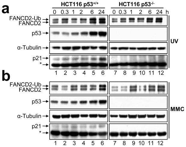
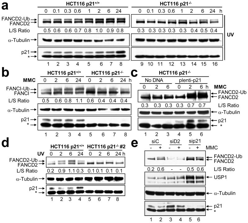
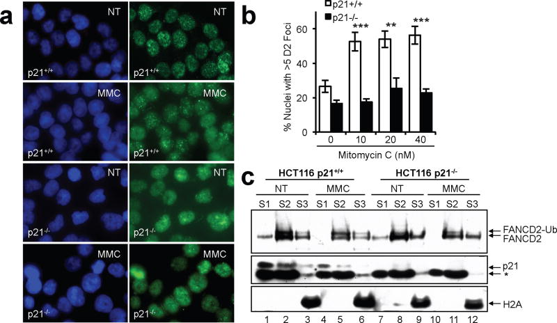
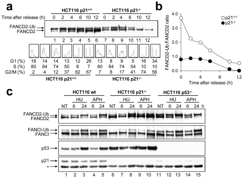
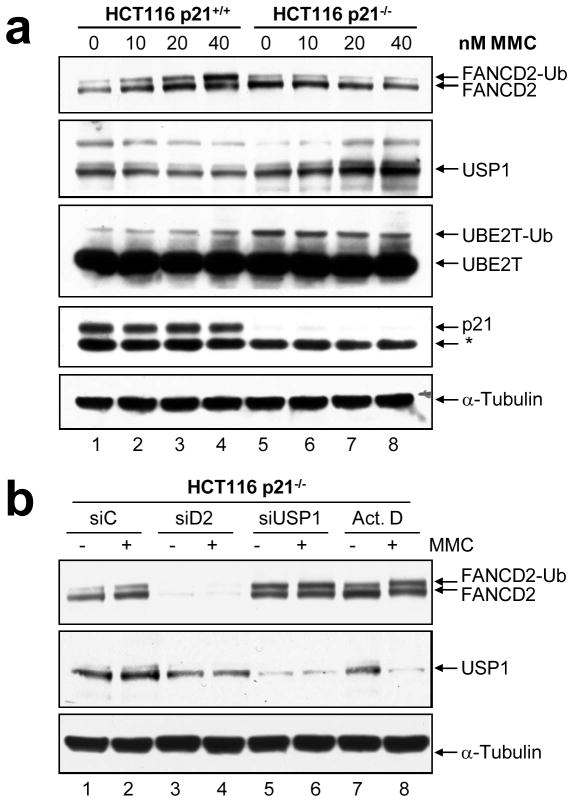
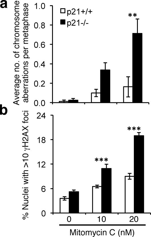
Fanconi anemia (FA) is a rare disease characterized by congenital defects, progressive bone marrow failure and heightened cancer susceptibility. The FA proteins, BRCA1 and FANCD1/BRCA2 function cooperatively in the FA-BRCA pathway to repair damaged DNA. Activation of the FA-BRCA pathway occurs via the monoubiquitination of the FANCD2 and FANCI proteins, targeting these proteins to discrete nuclear foci where they function in DNA repair. The cellular regulation of FANCD2/I monoubiquitination, however, remains poorly understood. In this study, we have examined the roles of the p53 tumor suppressor protein, as well as its downstream target, the p21(Cip1/Waf1) cyclin-dependent kinase inhibitor, in the regulation of the activation of the FA-BRCA pathway. We demonstrate that, in contrast to p53, p21 has a major role in the regulation of the activation of the FA-BRCA pathway: p21 promotes S-phase and DNA damage-inducible FANCD2/I monoubiquitination and nuclear foci formation. Several lines of evidence establish that this effect is not a consequence of a defective G1-S checkpoint or altered cell-cycle progression in the absence of p21. Instead, we demonstrate that p21 is required for the transcriptional repression of the USP1 deubiquitinating enzyme upon exposure to DNA-damaging agents. In the absence of p21, persistent USP1 expression precludes the DNA damage-inducible accumulation of monoubiquitinated FANCD2 and FANCI. Consequently, p21(-/-) cells exhibit increased levels of mitomycin C-inducible complex chromosomal aberrations and elevated γH2AX nuclear foci formation. Our results demonstrate that p21 has a critical role in the regulation of the activation of the FA-BRCA pathway and suggest a broader role for p21 in the orchestration of DNA repair processes following exposure to DNA crosslinking agents.


Conflict of interest statement

Conflict of interest

There are no competing financial interests in relation to the work described.

Figures

Figure 1
Figure 1
FANCD2 monoubiquitination is p53-independent. (a) HCT116 p53+/+ and p53−/− cells were treated with 20 J/m2 UV-C irradiation (UV) and 100 nM mitomycin C (MMC) for 24 h (b). Whole-cell lysates were prepared at the indicated time points, resolved, and immunoblotted with anti-FANCD2, anti-p53, anti-p21, and anti-α-tubulin antibodies. *, non-specific band.
Figure 2
Figure 2
p21 is required for DNA damage-inducible FANCD2 and FANCI monoubiquitination. (a) HCT116 p21+/+ and p21−/− cells were treated with 20 J/m2 UV-C irradiation (UV) and 100 nM mitomycin C (MMC) for 24 h (b). Whole cell lysates were prepared at the indicated time points, resolved, and immunoblotted with anti-FANCD2, anti-FANCI, anti-α-tubulin, and anti-p21 antibodies. (c) Transfection of HCT116 p21−/− cells with pLenti6.2-p21 restores DNA damage-induced FANCD2 monoubiquitination. Cells were treated with 100 nM MMC. (d) A second independently isolated HCT116 p21−/− clone demonstrates markedly attenuated DNA damage-inducible FANCD2 monoubiquitination following treatment with 20 J/m2 UV-C irradiation. *, non-specific band. (e) U2OS cells were transfected with control non-targeting (siC), FANCD2 (siD2), or p21 (sip21) siRNAs. Seventy-two hours post-transfection, cells were untreated (−) or treated (+) with 200 nM mitomycin C (MMC). Whole-cell lysates were prepared 6 h later, resolved, and immunoblotted with anti-FANCD2, anti-USP1, anti-p21, and anti-α-tubulin antibodies. The L/S ratio is the ratio of FANCD2-Ub (long isoform) to FANCD2 (short isoform).
Figure 3
Figure 3
In the absence of p21 FANCD2 localizes to chromatin yet fails to assemble into DNA damage-inducible nuclear foci. (a) HCT116 p21+/+ and p21−/− cells were untreated (NT) or exposed to mitomycin C (MMC) for 16 h. Cells were fixed and immunostained with a polyclonal antibody against FANCD2 (green) and counterstained with DAPI (blue). (b) The percentage of nuclei with > 5 FANCD2 foci were counted and plotted. At least 300 nuclei were scored per cell line and treatment. Error bars represent the standard errors of the means. **, p < 0.01; ***, p < 0.001. (c) Cells were incubated in the absence and presence of 60 nM MMC for 18 h, fractionated into cytoplasmic (S1), soluble nuclear (S2), and chromatin-associated fractions (S3), and immunoblotted with antibodies to FANCD2, p21, and H2A. *, non-specific band.
Figure 4
Figure 4
p21-dependent FANCD2/I monoubiquitination is cell cycle independent. (a) HCT116 p21+/+ and p21−/− cells were synchronized via double thymidine block, released into thymidine-free media and pellets collected for immunoblotting with anti-FANCD2 (top panel) and FACS analysis (bottom panel) at the indicated time points. (b) Band intensities from (a) were quantified using ImageJ software and plotted. (c) HCT116 wild type, p21−/− and p53−/− cells were untreated (NT) or treated with hydroxyurea (HU) and aphidicolin (APH), whole cell lysates were prepared, and resolved proteins immunoblotted with anti-FANCD2, anti-FANCI, anti-p53, and anti-p21 antibodies. *, non-specific band. For (b), while the band intensities for a single experiment are shown, this experiment was repeated multiple times with very similar findings.
Figure 5
Figure 5
Altered regulation of the USP1 and UBE2T enzymes in p21-null cells. (a) HCT116 p21+/+ and p21−/− cells were incubated in the absence or presence of the indicated concentrations of mitomycin C (MMC) for 24 h, whole-cell lysates prepared, and FANCD2, USP1, UBE2T, p21, and α-tubulin protein expression was analyzed by immunoblotting. (b) HCT116 p21−/− cells were transfected with control (non-targeting) (siC), FANCD2 (siD2), and USP1 (siUSP1) siRNAs. Seventy-two hours later, cells were untreated (−) or treated (+) with 40 nM mitomycin C (MMC) for 24 h, in the presence or absence of 4 μM actinomycin D (Act. D). Whole-cell lysates were prepared, and FANCD2, USP1, and α-tubulin protein expression was analyzed by immunoblotting.
Figure 6
Figure 6
p21−/− cells display elevated mitomycin C-inducible chromosome aberrations and γH2AX nuclear foci formation. (a) HCT116 p21+/+ and p21−/− cells were incubated in the absence and presence of 10 and 20 nM MMC for one cell cycle, metaphase spreads were prepared and chromosome aberrations, including chromosome and chromatid gaps and breaks and radial formations, were scored. At least 100 metaphases were scored per treatment. (b) HCT116 p21+/+ and p21−/− cells were treated as for (a) and the number of nuclei displaying >10 discrete γH2AX foci recorded. Error bars represent standard errors of the means and all experiments were performed in triplicate with similar findings. **, p < 0.01; ***, p < 0.001

References

    1. Abbas T, Dutta A. p21 in cancer: intricate networks and multiple activities. Nat Rev Cancer. 2009;9:400–14. - PMC - PubMed
    1. Abbas T, Sivaprasad U, Terai K, Amador V, Pagano M, Dutta A. PCNA-dependent regulation of p21 ubiquitylation and degradation via the CRL4Cdt2 ubiquitin ligase complex. Genes Dev. 2008;22:2496–506. - PMC - PubMed
    1. Abukhdeir AM, Park BH. P21 and p27: roles in carcinogenesis and drug resistance. Expert Rev Mol Med. 2008;10:e19. - PMC - PubMed
    1. Adamo A, Collis SJ, Adelman CA, Silva N, Horejsi Z, Ward JD, et al. Preventing nonhomologous end joining suppresses DNA repair defects of Fanconi anemia. Mol Cell. 2010;39:25–35. - PubMed
    1. Alpi A, Langevin F, Mosedale G, Machida YJ, Dutta A, Patel KJ. UBE2T, the Fanconi anemia core complex, and FANCD2 are recruited independently to chromatin: a basis for the regulation of FANCD2 monoubiquitination. Mol Cell Biol. 2007;27:8421–30. - PMC - PubMed

Publication types

MeSH terms