. 2012 Jan 12;31(2):149-60.
doi: 10.1038/onc.2011.222. Epub 2011 Jun 20.

Stem cell marker (Nanog) and Stat-3 signaling promote MicroRNA-21 expression and chemoresistance in hyaluronan/CD44-activated head and neck squamous cell carcinoma cells

Affiliations

Stem cell marker (Nanog) and Stat-3 signaling promote MicroRNA-21 expression and chemoresistance in hyaluronan/CD44-activated head and neck squamous cell carcinoma cells

L Y W Bourguignon et al. Oncogene. .

Abstract

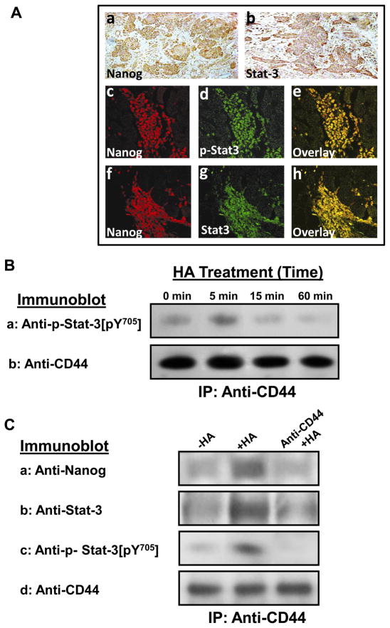
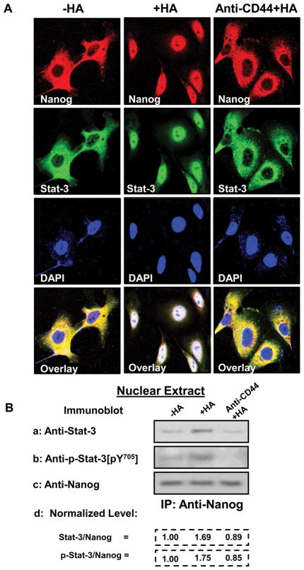
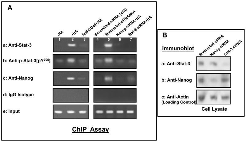
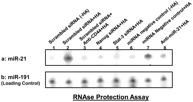
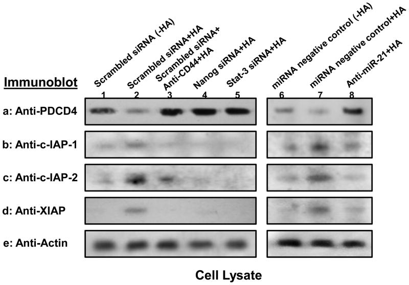
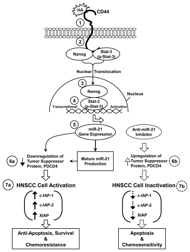
MicroRNAs are often associated with the pathogenesis of many cancers, including head and neck squamous cell carcinoma (HNSCC). In particular, microRNA-21 (miR-21) appears to have a critical role in tumor cell survival, chemoresistance and HNSCC progression. In this study, we investigated matrix hyaluronan (HA)-induced CD44 (a primary HA receptor) interaction with the stem cell markers, Nanog and Stat-3, in HNSCC cells (HSC-3 cells). Our results indicate that HA binding to CD44 promotes Nanog-Stat-3 (also tyrosine phosphorylated Stat-3) complex formation, nuclear translocation and transcriptional activation. Further analyses reveal that miR-21 is controlled by an upstream promoter containing Stat-3 binding site(s), while chromatin immunoprecipitation assays demonstrate that stimulation of miR-21 expression by HA/CD44 signaling is Nanog/Stat-3-dependent in HNSCC cells. This process results in a decrease of a tumor suppressor protein (PDCD4), and an upregulation of i nhibitors of the apoptosis family of proteins (IAPs) as well as chemoresistance in HSC-3 cells. Treatment of HSC-3 cells with Nanog- and/or Stat-3-specific small interfering RNAs effectively blocks HA-mediated Nanog-Stat-3 signaling events, abrogates miR-21 production and increases PDCD4 expression. Subsequently, this Nanog-Stat-3 signaling inhibition causes downregulation of survival protein (IAP) expression and enhancement of chemosensitivity. To further evaluate the role of miR-21 in tumor cell-specific functions, HSC-3 cells were also transfected with a specific anti-miR-21 inhibitor in order to silence miR-21 expression and block its target functions. Our results demonstrate that anti-miR-21 inhibitor not only upregulates PDCD4 expression but also decreases IAP expression and enhances chemosensitivity in HA-treated HNSCC cells. Together, these findings indicate that the HA-induced CD44 interaction with Nanog and Stat-3 has a pivotal role in miR-21 production leading to PDCD4 reduction, IAP upregulation and chemoresistance in HNSCC cells. This novel Nanog/Stat-3 signaling pathway-specific mechanism involved in miR-21 production is significant for the formation of future intervention strategies in the treatment of HA/CD44-activated HNSCC.


Conflict of interest statement

CONFLICT OF INTEREST: All authors declare no conflict of interest.

Figures

Fig. 1
Fig. 1. Analyses of Nanog and Stat-3 expression in HNSCC patient samples and HSC-3 cells
A: Detection of Nanog and Stat-3 expression in head and neck squamous cell carcinoma (HNSCC) primary tumors. A panel of immune-reagents including rabbit anti-Nanog antibody, rabbit anti-Stat-3 antibody or rabbit anti-phospho-Stat-3 (pY[705]) antibody were used to examine the expression of both Nanog and Stat-3 in HNSCC primary tumors using immuno-peroxidase staining (a and b) or co-localization of Nanog with phosphorylated Stat-3 (p-Stat-3) (c-e) and total stat-3 (f-h) using double immunofluorescence staining as described in the Materials and Methods. B: Time course analyses of HA/CD44-induced Stat-3 tyrosine phosphorylation: Cell lysates were prepared from HSC-3 treated with HA (50μg/ml) for various time intervals (e.g., 0, 5, 10, 15 and 60min) followed by anti-CD44-immunoprecipitation followed by immunoblotting with anti-phospho-Stat-3 [pY705] antibody or anti-CD44 antibody (as a loading control). C: Detection of Nanog and Stat-3 (or phosphorylated Stat-3) in the CD44 complex by anti-CD44-immunoprecipitation followed by immunoblotting with anti-Nanog antibody (a) or anti-Stat-3 antibody (b) or anti-phospho-Stat-3 [pY705] antibody (c) or reblotting with anti-CD44 (d) as a loading control using HSC-3 cells treated with no HA or with HA (50μg/ml) for 5min or pretreated with anti-CD44 antibody for 1h followed by 5min HA (50μg/ml) addition.
Fig. 2
Fig. 2. Immunocytochemical and biochemical analyses of HA/CD44-induced Nanog and Stat-3 nuclear translocation in HSC-3 cells
A: Immunostaining of Nanog [using Texas Red-labeled anti-Nanog (red color) and Stat-3 using FITC-labeled anti-Stat-3 (green color)]: HSC-3 cells (untreated or pretreated with anti-CD44 antibody for 1h) were incubated with HA (50μg/ml) (or without HA) for 15min at 37°C and fixed by 2% paraformaldehyde. Subsequently, these cells were rendered permeable by ethanol treatment and immuno-stained with Texas Red-labeled anti-Nanog (red color) or FITC-labeled anti-Stat-3 (green color) and DAPI (a nuclear marker) (c) as described in the Materials and Methods. B: Analyses of HA/CD44-induced Nanog and Stat-3 complex formation in the nuclear fraction: HSC-3 cells (untreated or pretreated with anti-CD44 antibody for 1h) were incubated with HA (50μg/ml) (or without HA) for 15min at 37°C. Nuclear fractions of these cells were then prepared by anti-Nanog-immunoprecipitation followed by immunoblotting with anti-Stat-3 antibody (a) or anti-phospho-Stat-3 (pY705) (b) or reblotting with anti-Nanog (c) as a loading control as described in the Materials and Methods [untreated cells (-HA); cells treated with HA (+HA) for 15min; lane 3: cells pretreated with anti-CD44 antibody for 1h plus 15min HA addition (anti-CD44 +HA)]. [The ratio of Stat-3 or phosphorylated Stat-3 and Nanog (the loading control) (d) was determined by densitometry, and the levels were normalized to untreated (no HA treatment) value (designated as 1.00); the values expressed represent an average of triplicate determination of 3 experiments with an SD of less than 5%].
Fig. 3
Fig. 3. Interaction between Nanog, Stat-3 and the upstream promoter/enhancer region of miR-21 promoter in HSC-3 cells
A: In vivo binding of Nanog and Stat-3 (or phosphorylated Stat-3) to the miR-21 upstream promoter/enhancer region in HSC-3 cells: ChIP assay was performed in HSC-3 cells following protocols described in the Materials and Methods using the Stat-3 binding site-containing miR-21 promoter (upstream promoter/enhancer region)-specific primers by PCR. Identical volumes from the final precipitated materials were used for the PCR reactions [untreated cells (lane 1); cells treated with HA for 30min (lane 2); cells pretreated with anti-CD44 antibody for 1h plus 30min HA addition (lane 3); cells pretreated with scrambled siRNA with no HA (lane 4); cells pretreated with scrambled siRNA plus 30min HA addition (lane 5); cells pretreated with Nanog siRNA plus 30min HA addition (lane 6); cells pretreated with Stat-3 siRNA plus 30min HA addition) (lane 7). (a: anti-Stat-3-mediated immunoprecipitated material; b: anti-phospho-Stat-3 (pY705)-mediated immunoprecipitated material; c: anti-Nanog-mediated immunoprecipitated material; d: IgG isotype control-mediated precipitated material; e: total input materials). B: Verification of the specificity of Nanog siRNA or Stat-3 siRNA used in the study. Cell lysates isolated from HSC-3 cells [transfected with Nanog siRNA or Stat-3 siRNA-target or scrambled siRNA] were solubilized by 1% Nonidet P-40 (NP-40) buffer followed by immunoblotting with anti-Stat-3 antibody (a), anti-Nanog antibody (b) or anti-actin (a loading control) (c).
Fig. 4
Fig. 4. Detection of HA/CD44-induced miR-21 production in HSC-3 cells
Detection of miR-21 in HSC-3 cells using RNase protection assay as described in the Materials and methods. A: Autoradiogram of miR-21 detected in HSC-3 cells incubated with scrambled sequence siRNA [without HA (lane 1) or with 2h HA treatment (lane 2) or pretreated with anti-CD44 antibody for 1h followed by HA addition for 2h (lane 3) or incubated with Nanog siRNA plus 2h HA treatment (lane 4) or incubated with Stat-3 siRNA plus 2h HA treatment (lane 5) or incubated with miRNA-negative control [without HA (lane 6) or with 2h HA treatment (lane 7)] or incubated with an anti-miR-21 inhibitor plus 2h HA treatment (lane 8). (Autoradiogram of miR-191 in each gel lane was used as a loading control).
Fig. 5
Fig. 5. Analyses of HA/CD44-mediated PDCD4 and IAP expression in HSC-3 cells
Detection of HA/CD44-induced PDCD4 and IAP (cIAP-1, cIAP-2 and XIAP) expression in HSC-3 cells was performed by solubilizing cells with 1% Nonidet P-40 (NP-40) buffer followed by immunobloting with anti-PDCD4 antibody or anti-cIAP-1 antibody or anti-cIAP-2 antibody or anti-XIAP antibody, respectively as described in the Materials and Methods. First, cell lysates were prepared from HSC-3 cells treated with scrambled sequence siRNA [without HA (lane 1) or with HA for 24h (lane 2)] or treated with anti-CD44 antibody for 1h followed by 24h HA addition (lane 3) or treated with Nanog siRNA plus HA for 24h (lane 4) or treated with Stat-3 siRNA plus HA for 24h (lane 5) or treated with miRNA-negative control [without HA (lane 6) or with HA for 24h (lane 7)] or treated with anti-miR-21 inhibitor plus HA for 24h (lane 8). These samples were then immunoblotted with anti-PDCD4 antibody (a) or anti-cIAP-1 antibody (b) or anti-cIAP-2 antibody (c) or anti-XIAP antibody (d), respectively. The amount of actin detected by anti-actin-mediated immunoblot (e) in each gel lane was used as a loading control.
Fig. 6
Fig. 6. A proposed model for HA/CD44-mediated Nanog and Stat-3 signaling in the regulation of miRNA-21 production, oncogenesis and chemoresistence in HNSCC cells
The binding of HA to CD44 (step 1) promotes a complex formation between Nanog/Stat-3 (also tyrosine phosphorylated Stat-3, p-Stat-3) and CD44 (step 2). Both Nanog and Stat-3 (p-Stat-3) then translocate from the cytosol to the nucleus and interacts with the Stat-3-binding sites located at the upstream promoter/enhancer region of miR-21 (step 4), resulting in miR-21 gene expression and mature miR-21 production (step 5). The resultant miR-21 then functions to downregulate the tumor suppressor protein (PDCD4) (step 6a, indicated by the arrow with solid lines) and promote HNSCC activation leading to IAP (c-IAP-1, c-IAP-2 and XIAP) expression, HNSCC cell anti-apoptosis/survival and chemoresistance (step 7a, indicated by the arrow with solid lines). In direct contrast, treatment of HNSCC cells with an anti-miR-21 inhibitor induces tumor suppressor protein (PDCD4) upregulation (step 6b, indicated by the arrow with dash lines). Subsequently, these changes result in HNSCC cell inactivation including inhibition of IAP (c-IAP-1, c-IAP-2 and XIAP) expression (Fig. 7b, indicated by the arrow with dash lines), stimulation of apoptosis and enhancement of chemosensitivity in HNSCC cells. This newly-discovered Nanog/Stat-3 signaling pathway leading to miR-21 functioning should provide important drug targets for sensitizing tumor cell apoptosis and overcome chemoresistance in HA/CD44-activated head and neck cancer cells.

Comment in

Similar articles

Cited by

References

    1. Asangani IA, Rasheed SAK, Nikolova DA, Leupold JH, Colburn NH, Post S, et al. Allgayer H. MicroRNA-21 (miR-21) post-transcriptionally downregulates tumor suppressor Pdcd4 and stimulates invasion, intravasation and metastasis in colorectal cancer. Oncogene. 2008;27:2128–2136. - PubMed
    1. Bourguignon LY. Hyaluronan-mediated CD44 activation of RhoGTPase signaling and cytoskeleton function promotes tumor progression. Semin Cancer Biol. 2008;18:251–259. - PMC - PubMed
    1. Bourguignon LY, Peyrollier K, Xia W, Gilad E. Hyaluronan-CD44 interaction activates stem cell marker Nanog, Stat-3-mediated MDR1 gene expression, and ankyrin-regulated multidrug efflux in breast and ovarian tumor cells. J Biol Chem. 2008;283:17635–17651. - PMC - PubMed
    1. Bourguignon LY, Spevak CC, Wong G, Xia W, Gilad E. Hyaluronan-CD44 interaction with PKCε promotes oncogenic signaling by the stem cell marker, Nanog and the production of microRNA-21 leading to downregulation of the tumor suppressor protein, PDCD4, anti-apoptosis and chemotherapy resistance in breast tumor cells. J Biol Chem. 2009a;284:26533–26546. - PMC - PubMed
    1. Bourguignon LY, Xia W, Wong G. Hyaluronan-mediated CD44 interaction with p300 and SIRT1 regulates beta-catenin signaling and NFkappaB-specific transcription activity leading to MDR1 and Bcl-xL gene expression and chemoresistance in breast tumor cells. J Biol Chem. 2009b;284:2657–2671. - PMC - PubMed

Publication types

MeSH terms