Review
. 2011 Jun 20;12(7):597-606.
doi: 10.1038/ni.2059.

T-bet in disease

Affiliations
Review

T-bet in disease

Vanja Lazarevic et al. Nat Immunol. .

Abstract

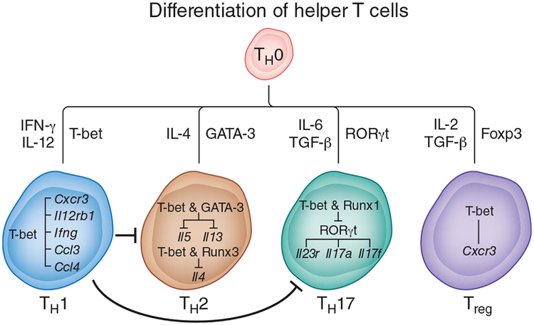
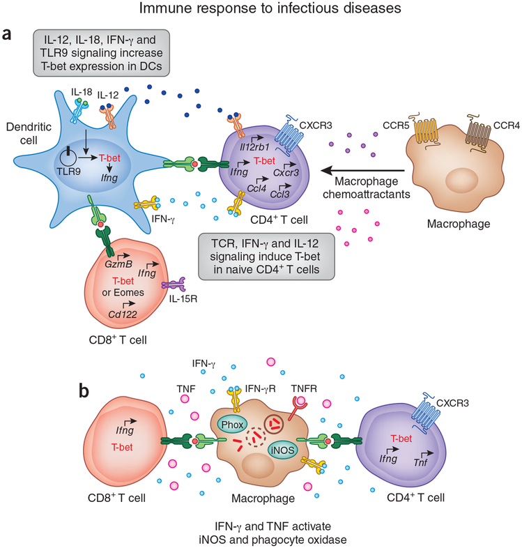
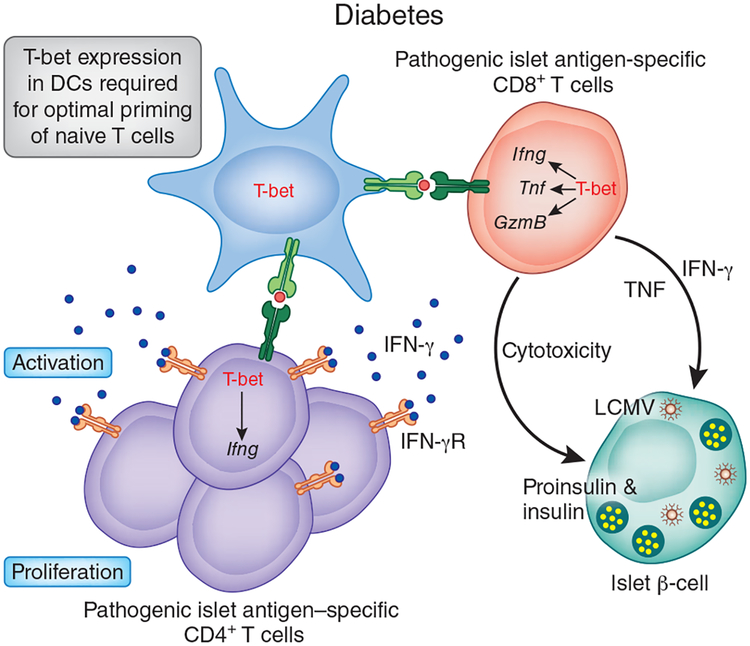
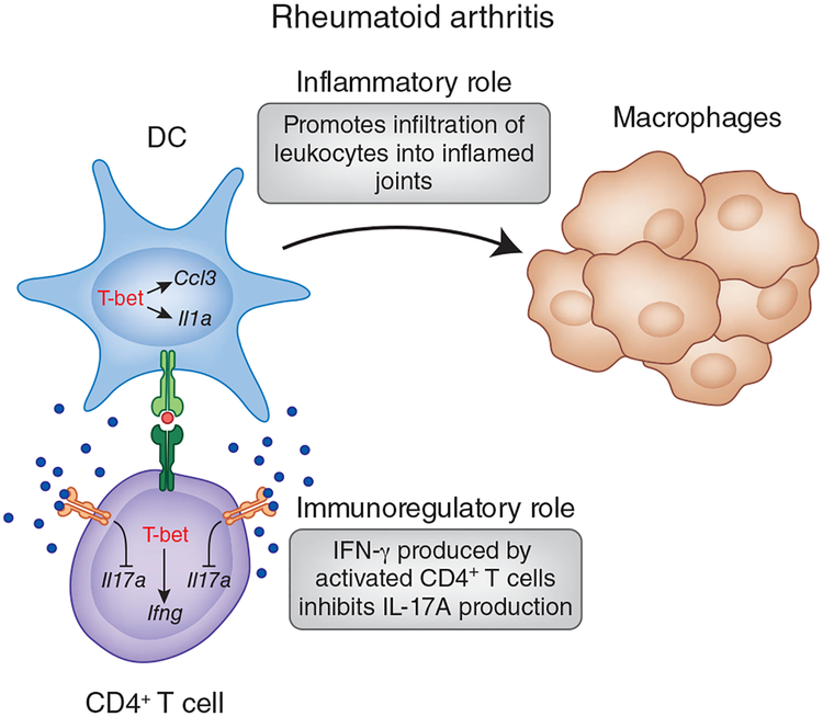
The activation of immune-defense mechanisms in response to a microbial attack must be robust and appropriately tailored to fight particular types of pathogens. Infection with intracellular microorganisms elicits a type 1 inflammatory response characterized by mobilization of T helper type 1 (T(H)1) cells to the site of infection, where they are responsible for the recruitment and activation of macrophages. At the center of the type 1 inflammatory response is the transcription factor T-bet, a critical regulator of the T(H)1 differentiation program. T-bet induces the production of interferon-γ (IFN-γ) and orchestrates the T(H)1 cell-migratory program by regulating the expression of chemokines and chemokine receptors. However, tight regulation of the type 1 inflammatory response is essential for the prevention of immunopathology and the development of organ-specific autoimmunity. In this review, we discuss how T-bet expression drives autoaggressive and inflammatory processes and how its function in vivo must be delicately balanced to avoid disease.


Conflict of interest statement

COMPETING FINANCIAL INTERESTS

The authors declare competing financial interests: details accompany the full-text HTML version of the paper at http://www.nature.com/natureimmunology/.

Figures

Figure 1
Figure 1
Role of T-bet in the differentiation of helper T cells. When naive CD4+ T cells (TH0) are activated in the presence of IFN-γ and IL-12, they differentiate into the TH1 subset. The differentiation of TH1 cells is critically dependent on the transcription factor T-bet. The first wave of T-bet expression in CD4+ T cells is regulated by signaling via the TCR and IFN-γ. T-bet upregulates the gene encoding the IL-12 receptor β2 subunit (Il12rb1) and confers IL-12 responsiveness, which induces the second wave of sustained T-bet expression. T-bet promotes TH1 differentiation not onlyby upregulating Ifng but also by inducing the expression of genes encoding CXCR3 and chemokines responsible for the mobilization of leukocytes tothe site of inflammation. In addition to promoting the TH1 differentiation program, T-bet suppresses commitment to the TH2 or TH17 lineage. T-bet blocks TH2 differentiation by sequestering the TH2-specific transcription factor GATA-3 away from the Il5 and Il13 promoters. T-bet and Runx3 bind to the Il4 silencer and prevent Il4 expression. In developing TH17 cells, T-bet binds to Runx1 and blocks expression of the TH17 cell–specific transcription factor RORγt and consequently RORγt target genes (Il23r, Il17a and Il17f). In fully differentiated TH17 cells, T-bet expression is associated with the appearance of repressive epigenetic changes in the Rorc locus, which result in the repression of Rorc expression. In Treg cells, T-bet expression is required for upregulation of the gene encoding CXCR3 and for the recruitment of Treg cells to the site of inflammation. T-bet expression in Treg cells is also essential for their suppressive activity in the scurfy model of autoimmunity but not in most organ-specific inflammatory or autoimmune diseases.
Figure 2
Figure 2
Role of T-bet in immune response to pathogens. (a) DCs express T-bet in response to signaling via IL-12, IL-18, IFN-γ and Toll-like receptor 9 (TLR9). T-bet expression in DCs is required for activation of the TH1 differentiation program in naive CD4+ T cells. In concert with TCR signaling, IFN-γ and IL-12 derived from mature DCs induce T-bet expression in CD4+ T cells and initiate TH1 differentiation. T-bet regulates the expression of genes encoding CXCR3, CCL3 and CCL4 by TH1 cells. CXCR3 is required for the migration of TH1 cells, whereas CCL3 and CCL4 are responsible for the recruitment of myeloid cellsto the site of inflammation. T-bet and eomesodermin (Eomes) have redundant roles in regulating the effector transcriptional program in CD8+ T cells. Both T-bet and eomesodermin control IFN-γ production and expression of the genes encoding granzyme B (Gzmb) and CD122 (the IL-2 and IL-15 receptor (IL-15R) β-subunit; CD122) by CD8+ T cells. Hence, mice deficient in either T-bet or eomesodermin demonstrate partial loss of cytotoxicity or partial deficiency in cytokine production relative to that of mice lacking both genes. IFN-γ production and granzyme B expressionare essential in immunity to intracellular pathogens, whereas CD122 expression is required for IL-15 responsiveness and the maintenance of memory CD8+ T cell responses in vivo. (b) IFN-γ and TNF delivered by effector CD4+ and CD8+ T cells activate microbicidal mechanisms in infected macrophages by inducing expression of phagocyte oxidase (Phox) and inducible nitric oxide synthase (iNOS). Reactive oxygen and nitrogen species generated by these two enzymesare responsible for the destruction of intracellular microorganisms. IFN-γR, IFN-γ receptor; TNFR, TNF receptor.
Figure 3
Figure 3
Role of T-bet in the pathogenesis of autoimmune diabetes. T-bet-deficient nonobese diabetic mice are protected from developing type 1 diabetes because of defects in their innate and adaptive immune systems. The priming ability of T-bet-deficient DCs is diminished, which results in the activation of fewer autoreactive TH1 cells. Less cytokine production by T-bet-deficient TH1 cells, which also have defective migration to the pancreas, causes the overall low-grade inflammatory response in the target organwith minimal damage. T-bet expression in CD8+ T cells is required for their pathogenicity in the RIP-LCMV transgenic model of virus-induced type 1 diabetes. T-bet-deficient mice have many fewer CD8+ effector-memory cells that produce IFN-γ and TNF and also have poor migratory potential.
Figure 4
Figure 4
Role of T-bet in the pathogenesis of rheumatoid arthritis. T-bet-deficient mice are protected from developing passive collagen antibody-induced arthritis. In this model, T-bet expression in DCs is required for disease pathogenesis. T-bet-deficient DCs are not efficient antigen-presenting cells and activate TH1 cells poorly. In the absence of T-bet, DCs produce much less IL-1α and CCL3 and recruit fewer leukocytes to the joints. In contrast, the role of T-bet in CD4+ T cells is less straightforward. In certain models, such as collagen- or proteoglycan-induced arthritis, expression of T-bet and IFN-γ may have an immunomodulatory effect on the development of arthritis by constraining the magnitude of TH17 responses.

References

    1. Mosmann TR, Cherwinski H, Bond MW, Giedlin MA & Coffman RL Two types of murine helper T cell clone. I. Definition according to profiles of lymphokine activities and secreted proteins. J. Immunol 136, 2348–2357 (1986). - PubMed
    1. Szabo SJ et al. A novel transcription factor, T-bet, directs Th1 lineage commitment. Cell 100, 655–669 (2000). - PubMed
    1. Zheng W & Flavell RA The transcription factor GATA-3 is necessary and sufficient for Th2 cytokine gene expression in CD4 T cells. Cell 89, 587–596 (1997). - PubMed
    1. Harrington LE et al. Interleukin 17-producing CD4+ effector T cells develop via a lineage distinct from the T helper type 1 and 2 lineages. Nat. Immunol 6, 1123–1132 (2005). - PubMed
    1. Park H et al. A distinct lineage of CD4 T cells regulates tissue inflammation by producing interleukin 17. Nat. Immunol 6, 1133–1141 (2005). - PMC - PubMed

Publication types

Substances