. 2011 Jul;1(3):324-37.
doi: 10.1016/j.dcn.2011.03.004.

How grossed out are you? The neural bases of emotion regulation from childhood to adolescence

Affiliations

How grossed out are you? The neural bases of emotion regulation from childhood to adolescence

Naomi B Pitskel et al. Dev Cogn Neurosci. 2011 Jul.

Abstract

The ability to regulate one's emotions is critical to mental health and well-being, and is impaired in a wide range of psychopathologies, some of which initially manifest in childhood or adolescence. Cognitive reappraisal is a particular approach to emotion regulation frequently utilized in behavioral psychotherapies. Despite a wealth of research on cognitive reappraisal in adults, little is known about the developmental trajectory of brain mechanisms subserving this form of emotion regulation in children. In this functional magnetic resonance imaging study, we asked children and adolescents to up-and down-regulate their response to disgusting images, as the experience of disgust has been linked to anxiety disorders. We demonstrate distinct patterns of brain activation during successful up- and down-regulation of emotion, as well as an inverse correlation between activity in ventromedial prefrontal cortex (vmPFC) and limbic structures during down-regulation, suggestive of a potential regulatory role for vmPFC. Further, we show age-related effects on activity in PFC and amygdala. These findings have important clinical implications for the understanding of cognitive-based therapies in anxiety disorders in childhood and adolescence.

Keywords: cognitive reappraisal; development; emotion regulation; functional magnetic resonance imaging.


Figures

Fig. 1
Fig. 1
Mean affect ratings as a function of condition. Error bars represent standard error of the mean (SEM). Single asterisk reflects significance at a threshold of p < 0.01; double asterisk reflects significance at a threshold of p < 0.001.
Fig. 2
Fig. 2
Brain activation in each of the three contrasts look-gross > look-neutral, decrease-gross > look-gross, increase-gross > look-gross, as well as the conjunction of the two regulation contrasts. Orange indicates positive-going activation; blue negative-going activation. All activations are at a threshold of p < 0.05. Images are displayed in radiologic convention. Talairach coordinates displayed to the left apply to the first three columns; those displayed on the right apply to the right-most column.
Fig. 3
Fig. 3
Top: Activation modulated by decrease-gross > look-gross in regions that were responsive to disgusting images. Blue indicates negative-going activation. All activations are at a threshold of p < 0.05. Bottom: Functionally defined ROIs in the right insula (yellow) and left amygdala (orange). Images are displayed in radiologic convention. Bar graph: y-axis represents differences in beta weights between the conditions of interest (decrease − look and increase − look).
Fig. 4
Fig. 4
Activation inversely correlated with left amygdala and right insula activation in the contrast decrease-gross > look-gross. Orange indicates regions of inverse correlation with seed regions, depicted in blue. All activations are at a threshold of p < 0.05. Images are displayed in radiologic convention.
Fig. 5
Fig. 5
Activation correlated with age in each of the three contrasts look-gross > look-neutral, decrease-gross > look-gross, and increase-gross > look-gross. Orange indicates regions that positively correlate with age; blue indicates regions that negatively correlate with age. All activations are at a threshold of p < 0.05. Images are displayed in radiologic convention. The graph to the right demonstrates the age distribution of the participants.
Supplemental Fig. 1
Supplemental Fig. 1
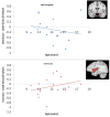
Top: Scatterplot of age versus difference β values for the contrast of decrease-gross > look-gross in the anatomically defined left amygdala. Bottom: Scatterplot of age versus difference β values for the contrast of increase-gross > look-gross in the anatomically defined left insula. The anatomical regions of interest are displayed in the upper right corner of each graph.

Similar articles

Cited by

References

    1. Aichhorn M., Perner J., Kronbichler M., Staffen W., Ladurner G. Do visual perspective tasks need theory of mind? Neuroimage. 2006;30:1059–1068. - PubMed
    1. Amaral D.G., Price J.L. Amygdalo-cortical projections in the monkey (Macaca fascicularis) J. Comp. Neurol. 1984;230:465–496. - PubMed
    1. Amstadter A. Emotion regulation and anxiety disorders. J. Anxiety Disord. 2008;22:211–221. - PMC - PubMed
    1. Banks S.J., Eddy K.T., Angstadt M., Nathan P.J., Phan K.L. Amygdala-frontal connectivity during emotion regulation. Soc. Cogn. Affect. Neurosci. 2007;2:303–312. - PMC - PubMed
    1. Barbas H. Anatomic basis of cognitive–emotional interactions in the primate prefrontal cortex. Neurosci. Biobehav. Rev. 1995;19:499–510. - PubMed

Publication types

LinkOut - more resources