. 2011 Jan;1(1):12-20.
doi: 10.4161/cl.1.1.13638.

Involvement of pigment globules containing multiple melanosomes in the transfer of melanosomes from melanocytes to keratinocytes

Affiliations

Involvement of pigment globules containing multiple melanosomes in the transfer of melanosomes from melanocytes to keratinocytes

Hideya Ando et al. Cell Logist. 2011 Jan.

Abstract

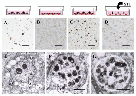
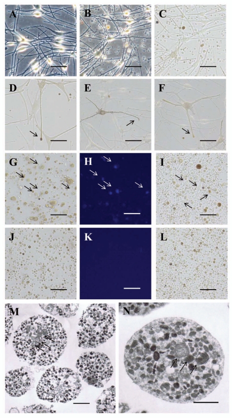
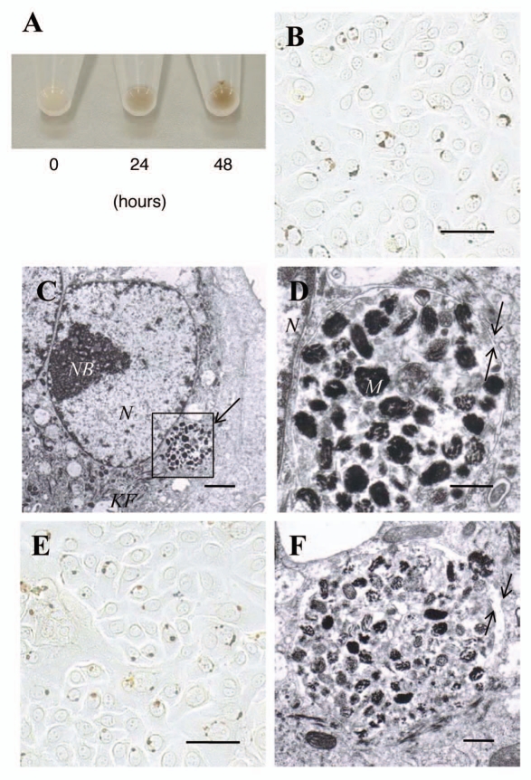
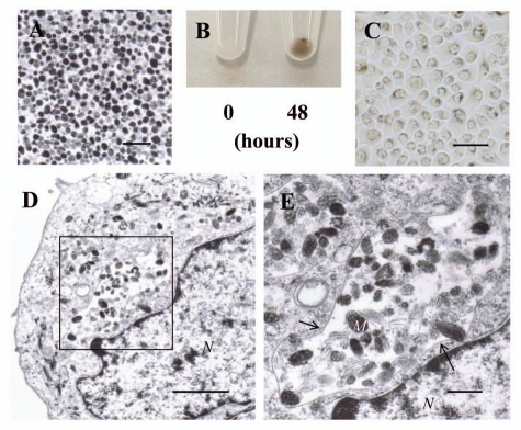
The mechanism of melanosome transfer from melanocytes to keratinocytes has not been fully clarified. We now show a route of melanosome transfer using co-cultures of normal human melanocytes and keratinocytes. Substantial levels of melanosome transfer were elicited in co-cultures of melanocytes and keratinocytes separated by a microporous membrane filter. The melanocyte dendrites penetrated into the keratinocyte layer through the filter and many pigment globules were observed in keratinocytes. Electron microscopic observations revealed that melanosomes incorporated in keratinocytes were packed in clusters enclosed by a double membrane. Numerous pigment globules budded off from melanocyte dendrites and were released into the culture medium. Those pigment globules were filled with multiple melanosomes and a few mitochondria but no nuclei. When those globules were added to the culture medium of keratinocytes, they were incorporated and showed double membrane-enclosed melano-phagolysosomes consistent with the structures obtained from the co-culture system. In contrast, when individual naked melanosomes isolated from melanocytes were added to keratinocytes, they were also phagocytosed by keratinocytes but were enclosed by a single membrane in a manner distinct from the co-culture system. These results suggest a novel mechanism of melanosome transfer, wherein melanosomes are packed in membrane globules that bud off from melanocyte dendrites, where they are released into the extracellular space and then phagocytosed by keratinocytes.


Figures

Figure 1
Figure 1
The microporous membrane filter-separated melanocyte and/or keratinocyte culture system. (A) Light microscopic image (bright field) of the bottom of a culture dish after growth of normal human melanocytes for 6 days. During the culture, there was a 1 µm-pore microporous membrane filter between the melanocytes and the bottom of the culture dish. After the membrane filter was removed, Fontana-Masson staining was performed. Many globules and a few segments (arrows) of melanocyte dendrites were dispersed. Scale bars in (A–D): 50 µm. (B) Light microscopic image (bright field) of the bottom of the culture dish after growth of normal human keratinocytes for 6 days. Fontana-Masson staining was performed to detect melanin but no staining was observed. (C) Light microscopic image (bright field) of the bottom of a culture dish after growth of normal human melanocytes and keratinocytes for 6 days. During the culture period, there was a 1 µm-pore microporous membrane filter between melanocytes and keratinocytes. Fontana-Masson staining was performed to detect melanin and single or clusters of pigment granules was/were observed closely associated with nuclei. (D) Light microscopic image (bright field) of the keratinocyte layer in the presence of 2 mg/mL STI, a known inhibitor of melanosome transfer, for 6 days. Fontana-Masson staining was performed and a decrease of melanosome transfer was observed. (E) Representative electron microscopic image of a normal human keratinocyte in C; the ingested melanosomes were packed and located in the perinuclear area (arrows). Scale bar: 2 µm. N: nuclei, NB: nuclear bodies. (F) Higher magnification image of the boxed region in E showing various stages of melanosomes or premelanosomes surrounded by a double membrane (arrows). Scale bar: 0.5 µm. M: representative melanosomes, N: nuclei. (G) Another electron microscopic image of ingested melanosomes in the co-culture system that shows an apparent double membrane (arrows) surrounding melanosomes. Scale bar: 0.5 µm. M: representative melanosomes.
Figure 2
Figure 2
Pigment globules containing multiple melanosomes released into the culture medium by normal human melanocytes. (A) Light microscopic image (phase contrast) of normal human melanocytes immediately after the culture medium was changed. Scale bars in (A–L): 50 µm. (B) Light microscopic image (phase contrast) of normal human melanocytes 24 hours after the culture medium was changed; note that numerous globules are present in the culture medium. (C) Bright field image of B; various degrees of pigmentation are observed in the globules. (D–F) Representative light microscopic images (bright field) of dendrites of normal human melanocytes. Pigment globules can be seen in the extended tip of a melanocyte dendrite (arrows). (G–I) Light microscopic images (G and I: bright field, H: fluorescence) of the culture medium of B after nuclear staining with DAPI (G and H) or incubated for 24 hours after re-seeding (I); many melanocytes (arrows in G and H indicate nuclei from the same positions, arrows in I indicate melanocyte cell bodies) and globules of various sizes are seen in the culture medium. (J–L) Light microscopic images (J and L: bright field, K: fluorescence) of the culture medium of B after filtration through an 8 µm-pore membrane filter; globules smaller than 8 µm diameter are seen but no melanocyte nuclei or melanocyte cell bodies are observed after DAPI staining (J and K) or after 24 hours of incubation (L). (M) Representative electron microscopic image of the purified pigment globules in (J–L); almost all globules contain multiple melanosomes and premelanosomes and a small number of mitochondria (arrows). Scale bar: 2 µm. (N) Higher magnification image of M; a large number of melanosomes and a small number of mitochondria (arrows) are surrounded by a single membrane. Scale bar: 1 µm.
Figure 3
Figure 3
Pigment globules containing multiple melanosomes derived from normal human melanocytes retain the capacity to transfer melanosomes to normal human keratinocytes. (A) Cell pellets of normal human keratinocytes incubated with the filtered pigment globules for 0, 24 or 48 hours; the pigmentation in the pellets after 24 and 48 hours of incubation is due to the pigment globules incorporated into the keratinocytes. (B) Light microscopic image (bright field) of normal human keratinocytes after incubation with the filtered pigment globules for 48 hours; Fontana-Masson staining revealed single or clustered pigment globules closely associated with nuclei. Scale bar: 50 µm. (C) Representative electron microscopic image of a normal human keratinocyte in (B); melanosomes and premelanosomes are packed and located in the perinuclear area. Scale bar: 2 µm. N: nuclei, NB: nuclear bodies, KF: keratin fibers. (D) Higher magnification image of the boxed region in C showing that the ingested melanosomes and premelanosomes are surrounded by a double membrane (arrows). Scale bar: 0.5 µm. M: representative melanosomes, N: nuclei. (E) Light microscopic image (bright field) of normal human keratinocytes after incubation with the globules or segments generated from melanocyte dendrites that had penetrated through the 1 µm membrane filter for 48 hours; Fontana-Masson staining revealed single or clustered pigment globules closely associated with nuclei. Scale bar: 50 µm. (F) Representative electron microscopic image of a normal human keratinocyte in E at higher magnification; melanosomes and premelanosomes are enclosed by a double membrane (arrows). Scale bar: 0.5 µm. M: representative melanosomes.
Figure 4
Figure 4
Uptake of individual naked melanosomes isolated from normal human melanocytes by normal human keratinocytes. (A) Electron microscopic image of isolated naked melanosomes. Scale bar: 1 µm. (B) Cell pellets of normal human keratinocytes incubated with isolated naked melanosomes for 0 or 48 hours. The pigmentation in the pellet after 48 hours of incubation is elicited by the naked melanosomes incorporated by normal human keratinocytes. (C) Light microscopic image (bright field) of normal human keratinocytes after incubation with isolated naked melanosomes for 48 hours; Fontana-Masson staining revealed that the dispersed pigment granules had accumulated in the perinuclear area. Scale bar: 50 µm. (D) Representative electron microscopic image of a normal human keratinocyte in (C); the melanosomes and premelanosomes are present in clusters and are enclosed by an amorphous structure membrane. Scale bar: 2 µm. N: nuclei. (E) Higher magnification image of the boxed region in (D) showing that the ingested melanosomes and premelanosomes are surrounded by a distorted single membrane (arrows). Scale bar: 0.5 µm. M: representative melanosomes, N: nuclei.
Figure 5
Figure 5
Scheme depicting traditional concepts and the new proposed melanosome transfer pathway. (A) Traditional concept showing direct inoculation of melanosomes into keratinocytes through nanotubular filopodia. (B) Traditional concept showing individual melanosome exocytosed from melanocytes and taken up by keratinocytes via membrane fusion-associated endocytosis (e) or phagocytosis (p). (C) Traditional concept showing cytophagocytosis of melanocyte dendrite tips by adjacent keratinocytes. (D) New concept showing numerous melanosomes packed in globules enclosed by the plasma membrane of the melanocyte, released into the extracellular space (into the culture medium) and finally being phagocytosed by normal human keratinocytes and then turned to melano-phagolysosomes enclosed by a double membrane. Color indications: melanocyte plasma membrane is blue, keratinocyte plasma membrane is red, and melanosomal membrane is green.
Figure 6
Figure 6
Photos of the originally established materials and methods. (A) Material for the microporous (1 µm-pore) membrane filter separating the melanocyte-keratinocyte co-culture system. The microporous membrane filter is placed between the culture dish (lower) and a top cover (upper). The material was finally placed on the 10 mm dish in order to hold and handle easily. (B) Method of filtering the culture medium of normal human melanocytes. A microporous (8 µm-pore) membrane filter was put on the dish and the culture medium was poured into the inner side of the microporous membrane filter. The liquid containing the pigment globules (diameter less than 8 µm) that penetrated the filter to remove melanocytes was collected slowly by capillary action.

References

    1. Hearing VJ. Biogenesis of pigment granules: a sensitive way to regulate melanocyte function. J Dermatol Sci. 2005;37:3–14. - PubMed
    1. Mottaz JH, Zelickson AS. Melanin transfer: a possible phagocytic process. J Invest Dermatol. 1967;49:605–610. - PubMed
    1. Yamamoto O, Bhawan J. Three modes of melanosome transfers in Caucasian facial skin: hypothesis based on an ultrastructural study. Pigment Cell Res. 1994;7:158–169. - PubMed
    1. Seiberg M. Keratinocyte-melanocyte interactions during melanosome transfer. Pigment Cell Res. 2001;14:236–242. - PubMed
    1. Scott G, Leopardi S, Printup S, Madden BC. Filopodia are conduits for melanosome transfer to keratinocytes. J Cell Sci. 2002;115:1441–1451. - PubMed

LinkOut - more resources