. 2011:122:82-92.

Regulation of renal urea transport by vasopressin

Affiliations

Regulation of renal urea transport by vasopressin

Jeff M Sands et al. Trans Am Clin Climatol Assoc. 2011.

Abstract

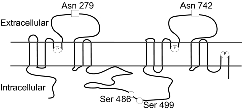
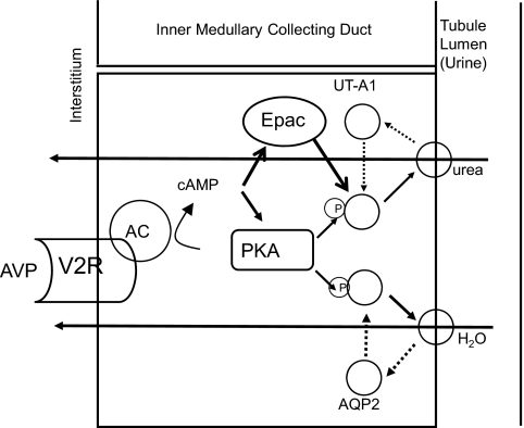
Terrestrial life would be miserable without the ability to concentrate urine. Production of concentrated urine requires complex interactions among the nephron segments and vasculature in the kidney medulla. In addition to water channels (aquaporins) and sodium transporters, urea transporters are critically important to the theories proposed to explain the physiologic processes occurring when urine is concentrated. Vasopressin (anti-diuretic hormone) is the key hormone regulating the production of concentrated urine. Vasopressin rapidly increases water and urea transport in the terminal inner medullary collecting duct (IMCD). Vasopressin rapidly increases urea permeability in the IMCD through increases in phosphorylation and apical plasma-membrane accumulation of the urea transporter A1 (UT-A1). Vasopressin acts through two cAMP-dependent signaling pathways in the IMCD: protein kinase A and exchange protein activated by cAMP Epac. Protein kinase A phosphorylates UT-A1 at serines 486 and 499. In summary, vasopressin regulates urea transport acutely by increasing UT-A1 phosphorylation and the apical plasma-membrane accumulation of UT-A1 through two cAMP-dependent pathways.


Conflict of interest statement

Potential Conflicts of Interest: None disclosed.

Figures

Fig. 1
Fig. 1
Diagram showing the locations of the chief urea transporters (UT-A1, UT-A2, UT-A3), aquaporins (AQP2-4), and sodium co-transporter (NKCC2) involved in urine concentration. The major regions of the kidney are indicated on the right. NaCl is actively reabsorbed across the thick ascending limb by the apical plasma-membrane Na-K-2C1 co-transporter (NKCC2). Water is reabsorbed across the descending limb segments by AQP1 water channels in both apical and basolateral plasma membranes. Water is reabsorbed across the apical plasma membrane of the entire collecting duct by AQP2 water channels when vasopressin is present. Water is reabsorbed across the basolateral plasma membrane by AQP3 water channels in the cortical and outer medullary collecting ducts and by both AQP3 and AQP4 water channels in the IMCD. Urea is concentrated within the collecting-duct lumen (by water reabsorption) until it reaches the terminal IMCD, where it is reabsorbed by the urea transporters UT-A1 and UT-A3.
Fig. 2
Fig. 2
Proposed membrane structure of UT-A1 (4, 5) showing the two established N-glycosylation sites (squares) (24) and the two established PKA phosphorylation sites (circles) (26). Also shown are the prolines that are putative β-turn sites (ovals containing P) (4, 5).
Fig. 3
Fig. 3
Urea and water reabsorption in principal cells of the inner medullary collecting duct. For water, vasopressin (AVP) binds to the V2-receptor (V2R) on the basolateral plasma membrane, activates adenylyl cyclase (AC), increases intracellular cyclic AMP (cAMP), and stimulates protein kinase A (PKA) activity. Cytoplasmic vesicles carrying the AQP2 water-channel protein are phosphorylated and inserted into the luminal membrane in response to vasopressin, thereby increasing the water permeability of this membrane (45). When vasopressin stimulation ends, water channels are retrieved by an endocytic process, and water permeability returns to its low basal rate. The process is similar for urea, except that cAMP stimulates both PKA and Epac, the exchange protein activated by cAMP (31, 33). UT-A1 is ubiquitinated and degraded in the proteasome (46). Although not shown, the basolateral plasma membrane contains AQP3 and AQP4 water channels and the UT-A3 urea transporter, thereby completing the transcellular pathways for water and urea reabsorption.

References

    1. Galluci E, Micelli S, Lippe C. Non-electrolyte permeability across thin lipid membranes. Arch Int Physiol Biochim. 1971;79:881–7. - PubMed
    1. Sands JM, Nonoguchi H, Knepper MA. Vasopressin effects on urea and H2O transport in inner medullary collecting duct subsegments. Am J Physiol. 1987;253:F823–32. - PubMed
    1. Wieth JO, Funder J, Gunn RB, Brahm J. Passive transport pathways for chloride and urea through the red cell membrane. In: Bolis K, Bloch K, Luria SE, Lynen F, editors. Comparative Biochemistry and Physiology of Transport. Amsterdam: Elsevier/North-Holland; 1974. pp. 317–37.
    1. Sands JM, Timmer RT, Gunn RB. Urea transporters in kidney and erythrocytes. Am J Physiol. 1997;273(3):F321–39. - PubMed
    1. Sands JM, Layton HE. The physiology of urinary concentration: an update. Semin Nephrol. 2009;29(3):178–95. - PMC - PubMed

Publication types

LinkOut - more resources