

Primary leiomyoma of the liver: accurate preoperative diagnosis on liver biopsy
- PMID: 21686574
- PMCID: PMC3027874
- DOI: 10.1136/bcr.09.2008.0898
Primary leiomyoma of the liver: accurate preoperative diagnosis on liver biopsy
Abstract
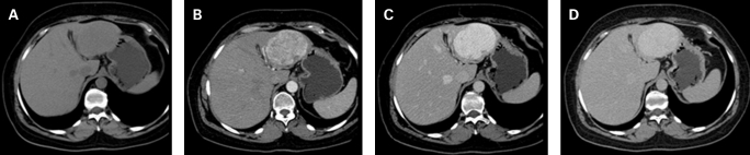
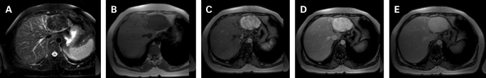
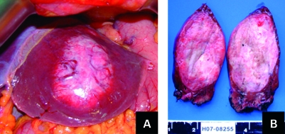
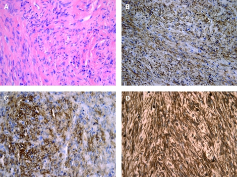
Primary leiomyoma of the liver is an exceptionally rare tumour in non-immunocompromised patients. Preoperative diagnosis of the lesion is difficult as complete imaging of this type of lesion is scarcely defined and preoperative biopsy was not the practice in previously reported cases. We report a voluminous primary leiomyoma of the liver occurring in a healthy middle-aged woman where a preoperative diagnosis was accurately achieved on biopsy. Because of its size, surgery was undertaken for exclusion of malignancy. A 16-month uneventful follow-up has been completed. We discuss the advantage of a preoperative diagnosis and propose that an imaging-guided liver biopsy should be undertaken, provided malignancy features are absent. This could prevent liver surgery merely for diagnostic purposes. Finally, we report imaging features that have not been previously described, namely on magnetic resonance imaging, which may provide an insight about the nature of this particular lesion and, advantageously, contribute toward a non-invasive diagnosis.
Figures
References
-
- Urizono Y, Ko S, Kanehiro H, et al. Primary leiomyoma of the liver: report of a case. Surg Today 2006; 36:629–32 - PubMed
-
- Kanazawa N, Izumi N, Tsuchiya K, et al. A case of primary leiomyoma of the liver in a patient without evidence of immunosuppression. Hepatol Res 2002; 24: 80–8 - PubMed
-
- Beuzen F, Roudie J, Moali I, et al. Léiomyome du foie: une tumeur benigne exceptionelle. Gastroenterol Clin Biol 2004; 28: 1169–72 - PubMed
-
- Guy CD, Yuan S, Ballo MS. Spindle-cell lesions of the liver: diagnosis by fine-needle aspiration biopsy. Diagn Cytopathol 2001; 25: 94–100 - PubMed
-
- Scoazec JY, Labadie M, Dumortier J, et al. Diagnostic des nodules hépatiques: techniques, démarche et principaux problèmes pratiques. Gastroenterol Clin Biol 2000; 24: 1095–103 - PubMed
LinkOut - more resources
Full Text Sources