Paediatric unilateral giant hydroureteronephrosis from idiopathic ureterovesical stricture: a case report
- PMID: 21686635
- PMCID: PMC3028028
- DOI: 10.1136/bcr.08.2008.0782
Paediatric unilateral giant hydroureteronephrosis from idiopathic ureterovesical stricture: a case report
Abstract
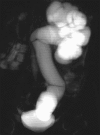
A congenital type of ureterovesical junction obstruction may be present in the fetus or at any stage during childhood, more commonly associated with urinary tract infections and other secondary causes. We present the case of a 6-year-old boy who suffered from colic and side pain, which was worsening monthly. He suffered from a giant hydroureteronephrosis resulting from idiopathic ureterovesical junction obstruction, with no clinical or laboratory signs of urinary tract infection or other secondary causes of obstruction. Indications for surgery were a decrease in kidney function (<40%) at scintigraphy, severe hydronephrosis (>30 mm), and the coexistence of symptoms (colic pain). After surgery, kidney function returned to almost completely normal. Unexpectedly an obstruction may become symptomatic late in infancy, especially in patients with normal prenatal ultrasound screening and postnatal life, as was the case for our patient in whom the only clinical sign was pain at flank.
Figures


References
-
- Hinds AC. Obstructive uropathy: considerations for the nephrology nurse (Continuing Education). Nephrol Nurs J 2004; 31: 166–74 - PubMed
-
- Oliveira EA, Diniz JS, Rabelo EA, et al. Primary megaureter detected by prenatal ultrasonography: conservative management and prolonged follow-up. Int Urol Nephrol 2000; 32: 13–8 - PubMed
-
- Becker A, Baum M. Obstructive uropathy. Early Hum Dev 2006; 82: 15–22 - PubMed
-
- Catalano C, Pavone P, Laghi A, et al. MR pyelography and conventional MR imaging in urinary tract obstruction. Acta Radiol 1999; 40: 198–202 - PubMed
LinkOut - more resources
Full Text Sources
