Dementia and leukoencephalopathy due to lymphomatosis cerebri
- PMID: 21686648
- PMCID: PMC3028137
- DOI: 10.1136/bcr.08.2008.0752
Dementia and leukoencephalopathy due to lymphomatosis cerebri
Abstract
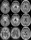
Lymphomatosis cerebri (LC) is a rare variant of primary central nervous system lymphoma (PCNSL). Clinically, the disease typically presents with a rapidly progressive dementia and unsteadiness of gait. Its presentation on cerebral MRI, which is characterised by diffuse leukoencephalopathy without contrast enhancement, often causes diagnostic confusion1 with suspected diagnoses ranging from Binswanger's disease to leukoencephalopathy or encephalomyelitis. Here we report a patient with subacute dementia and diffuse bilateral white matter changes in the cerebral hemispheres and additional involvement of the brainstem, basal ganglia and thalamus on MRI. Initially, she was considered to suffer from an autoimmune encephalitis, transiently responded to immunosuppression but then developed multiple solid appearing cerebral lymphomas.
Figures

References
-
- Rollins KE, Kleinschmidt-DeMasters BK, Corboy JR, et al. Lymphomatosis cerebri as a cause of white matter dementia. Hum Pathol 2005; 36: 282–90 - PubMed
-
- Kuker W, Nagele T, Korfel A, et al. Primary central nervous system lymphomas (PCNSL): MRI features at presentation in 100 patients. J Neurooncol 2005; 72: 169–77 - PubMed
-
- Bakshi R, Mazziotta JC, Mischel PS, et al. Lymphomatosis cerebri presenting as a rapidly progressive dementia: clinical, neuroimaging and pathologic findings. Dement Geriatr Cogn Disord 1999; 10: 152–7 - PubMed
-
- Matsumoto K, Kohmura E, Fujita T, et al. Recurrent primary central nervous system lymphoma mimicking neurodegenerative disease—an autopsy case report. Neurol Med Chir (Tokyo) 1995; 35: 360–3 - PubMed
-
- Mielke R, Kessler J, Szelies B, et al. Vascular dementia: perfusional and metabolic disturbances and effects of therapy. J Neural Transm Suppl 1996; 47: 183–91 - PubMed
LinkOut - more resources
Full Text Sources
