

A novel mitochondrial ATP8 gene mutation in a patient with apical hypertrophic cardiomyopathy and neuropathy
- PMID: 21686774
- PMCID: PMC3027703
- DOI: 10.1136/bcr.07.2008.0504
A novel mitochondrial ATP8 gene mutation in a patient with apical hypertrophic cardiomyopathy and neuropathy
Abstract
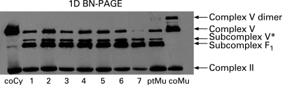
To identify the biochemical and molecular genetic defect in a 16-year-old patient presenting with apical hypertrophic cardiomyopathy and neuropathy suspected for a mitochondrial disorder.Measurement of the mitochondrial energy-generating system (MEGS) capacity in muscle and enzyme analysis in muscle and fibroblasts were performed. Relevant parts of the mitochondrial DNA were analysed by sequencing.A homoplasmic nonsense mutation m.8529G→A (p.Trp55X) was found in the mitochondrial ATP8 gene in the patient's fibroblasts and muscle tissue. Reduced complex V activity was measured in the patient's fibroblasts and muscle tissue, and was confirmed in cybrid clones containing patient-derived mitochondrial DNAWe describe the first pathogenic mutation in the mitochondrial ATP8 gene, resulting in an improper assembly and reduced activity of the complex V holoenzyme.
Figures


References
-
- Sperl W, Jesina P, Zeman J, et al. Deficiency of mitochondrial ATP synthase of nuclear genetic origin. Neuromuscul Disord 2006; 16: 821–9 - PubMed
-
- Nijtmans LG, Klement P, Houstek J, et al. Assembly of mitochondrial ATP synthase in cultured human cells: implications for mitochondrial diseases. Biochim Biophys Acta 1995; 1272: 190–8 - PubMed
-
- Capaldi RA, Aggler R, Turina P, et al. Coupling between catalytic sites and the proton channel in F1F0 type ATPases. Trends Biochem Sci 1994; 19: 284–9 - PubMed
-
- Boyer PD. The ATP synthase– a splendid molecular machine. Annu Rev Biochem 1997; 66: 717–49 - PubMed
-
- Noji H, Yasuda R, Yoshida M, et al. Direct observation of the rotation of F1-ATPase. Nature 1997; 386: 299–302 - PubMed
LinkOut - more resources
Full Text Sources
Molecular Biology Databases