

Enteropathy associated T cell lymphoma: common in coeliac disease
- PMID: 21686880
- PMCID: PMC3029034
- DOI: 10.1136/bcr.06.2008.0270
Enteropathy associated T cell lymphoma: common in coeliac disease
Abstract
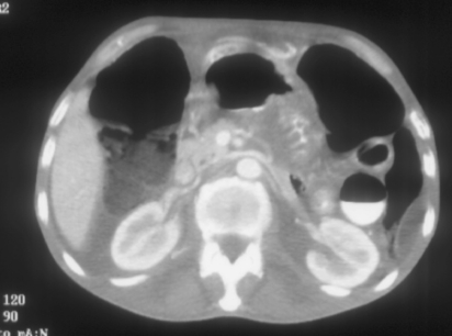
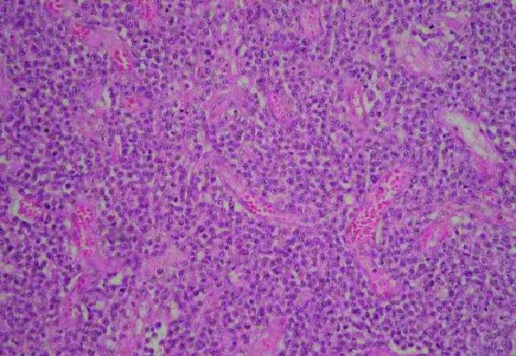
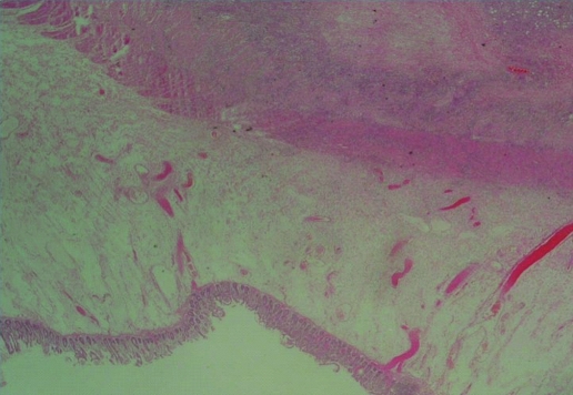
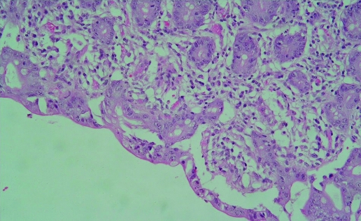
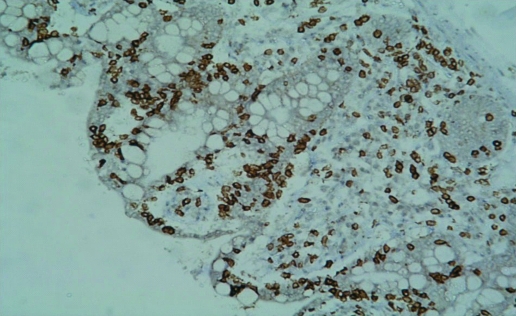
A 56-year-old male admitted with haematemesis and epigastric pain and severe weight loss on a background of coeliac disease. Computed tomography (CT) abdomen revealed a thickening of the mucosal folds of a short segment of jejunum. He deteriorated and had an exploratory laparotomy and bowel resection with side-side jejojejunal stapled anastomosis and extended right hemicolectomy and ileocolic anastomosis. Histology demonstrated multifocal high-grade malignant T cell lymphoma. Coeliac disease is a very common lifelong disorder. It is associated with osteoporosis, infertility, autoimmune disorders and increased risk of malignancy including an increased risk of non-Hodgkin's lymphoma (NHL) especially of the T cell type. Enteropathy-type T cell lymphoma is associated with a very poor prognosis. There is significant evidence that adherence to a gluten-free diet decreases the risk of developing enteropathy-type T cell lymphoma and helps to prevent development of autoimmune diseases, diabetes mellitus and osteoporosis in patients with coeliac disease.
Figures
Similar articles
-
Diffuse large B-cell lymphoma of the small intestine in a patient with refractory coeliac disease.Malays J Pathol. 2019 Apr;41(1):65-69. Malays J Pathol. 2019. PMID: 31025641
-
Clinical and pathological features of 14 non-Hodgkin's lymphomas associated with coeliac disease.Acta Clin Belg. 2004 May-Jun;59(3):143-51. doi: 10.1179/acb.2004.021. Acta Clin Belg. 2004. PMID: 15462511
-
Refractory sprue, coeliac disease, and enteropathy-associated T-cell lymphoma. French Coeliac Disease Study Group.Lancet. 2000 Jul 15;356(9225):203-8. doi: 10.1016/s0140-6736(00)02481-8. Lancet. 2000. PMID: 10963198
-
Complications of coeliac disease.Best Pract Res Clin Gastroenterol. 2015 Jun;29(3):451-8. doi: 10.1016/j.bpg.2015.05.005. Epub 2015 May 14. Best Pract Res Clin Gastroenterol. 2015. PMID: 26060109 Review.
-
Dermatitis herpetiformis: a cutaneous manifestation of coeliac disease.Ann Med. 2017 Feb;49(1):23-31. doi: 10.1080/07853890.2016.1222450. Epub 2016 Dec 14. Ann Med. 2017. PMID: 27499257 Review.
Cited by
-
An unusual presentation of EATL type 1: Emergency surgery due to life-threatening gastrointestinal bleeding.Int J Surg Case Rep. 2013;4(11):961-4. doi: 10.1016/j.ijscr.2013.08.007. Epub 2013 Aug 24. Int J Surg Case Rep. 2013. PMID: 24055918 Free PMC article.
References
-
- Dewar DH, Ciclitira PJ. Clinical features and diagnosis of celiac disease. Gastroenterology 2005; 128(1 Suppl): 19–24S - PubMed
-
- Catassi C, Ratsch IM, Fabiani E, et al. Coeliac disease in the year 2000: exploring the iceberg. Lancet 1994; 343: 200–3 - PubMed
-
- Green PH, Cellier C. Celiac disease. N Engl J Med 2007; 357: 1731–43 - PubMed
-
- Mäki M, Mustalahti K, Kokkonen J, et al. Prevalence of celiac disease among children in Finland. N Engl J Med 2003; 348: 2517–24 - PubMed
LinkOut - more resources
Full Text Sources