

doi: 10.1136/bcr.06.2008.0127.
Epub 2009 Apr 7.
Malignant neurilemoma with xeroderma pigmentosum
Affiliations
- PMID: 21686923
- PMCID: PMC3028145
- DOI: 10.1136/bcr.06.2008.0127
Item in Clipboard
Malignant neurilemoma with xeroderma pigmentosum
BMJ Case Rep.
2009.
Abstract
Xeroderma pigmentosum is a rare autosomal recessive disease characterised by hypersensitivity to sunlight, and is associated with a high incidence of skin cancer. We report a case of xeroderma pigmentosum with malignant neurilemoma in a 46-year-old woman which is unique due to its presentation, which was confirmed histopathologically.
Trial registration number: 31095.
Figures

The patient with a mass in the left orbit preoperatively. The pigmented lesion was raised 4–6 mm from the surface. She developed a firm mass in the interpalpebral zone of the left eye measuring 2.0×2.5 cm, which was attached to the palpebral conjunctiva and the eyelids.
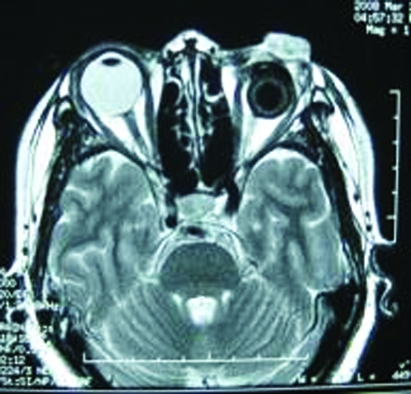
Magnetic resonance image disclosing a heterogeneous signal intensity on T2 weighted images.

Histological examination revealed a typical pattern of schwannoma consisting of Antoni type B. Histopathologically, the tumour tissue consists of so-called Antoni A and B type cells. Type A tissue shows densely packed, elongated spindle cells, while type B tissue has a more myxoid consistency. Haematoxylin and eosin; original magnification ×20.
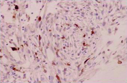
S-100 protein is positive in immunochemistry staining. Immunohistochemically, the tumour cells were partly positive for S-100. Original magnification ×200.

The mass in the left orbit was dissected 2 weeks postoperatively.
References
-
- Norgauer J, Idzko M, Panther E, et al. Xeroderma pigmentosum. Eur J Dermatol 2003; 13: 4–9 - PubMed
-
- Hebra F, Kaposi M. On diseases of the skin including exanthemata. New Sydenham Soc 1874; 61: 252–8
-
- Mseddi M, Sellami D, Elloumi Y, et al. Ophthalmologic manifestations of the xeroderma pigmentosum. Tunis Med 2006; 84: 542–4 - PubMed
-
- Yamashiro S, Nagahiro S, Mimata C, et al. Malignant trigeminal schwannoma associated with xeroderma pigmentosum—case report. Neurol Med Chir (Tokyo) 1994; 34: 817–20 - PubMed
-
- Nakamura T, Ono T, Yoshimura K, et al. Malignant schwannoma associated with xeroderma pigmentosum in a patient belonging to complementation group D. J Am Acad Dermatol 1991; 25(2pt 2): 349–53 - PubMed
LinkOut - more resources
Full Text Sources