. 2011 Feb 14:2:23.
doi: 10.3389/fmicb.2011.00023. eCollection 2011.

Molecular Characterization of Exploitation of the Polyubiquitination and Farnesylation Machineries of Dictyostelium Discoideum by the AnkB F-Box Effector of Legionella Pneumophila

Affiliations

Molecular Characterization of Exploitation of the Polyubiquitination and Farnesylation Machineries of Dictyostelium Discoideum by the AnkB F-Box Effector of Legionella Pneumophila

Tasneem Al-Quadan et al. Front Microbiol. .

Abstract

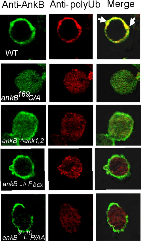
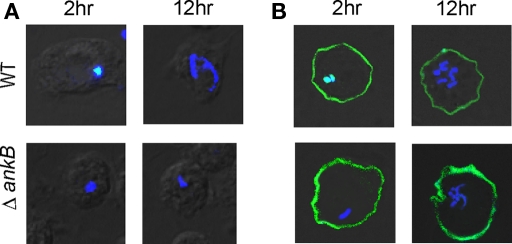
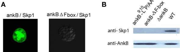
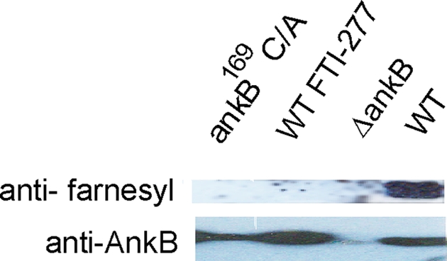
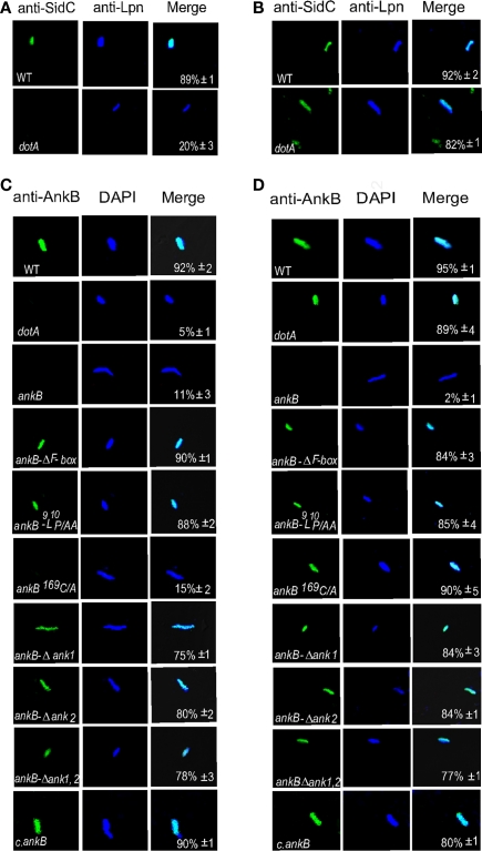
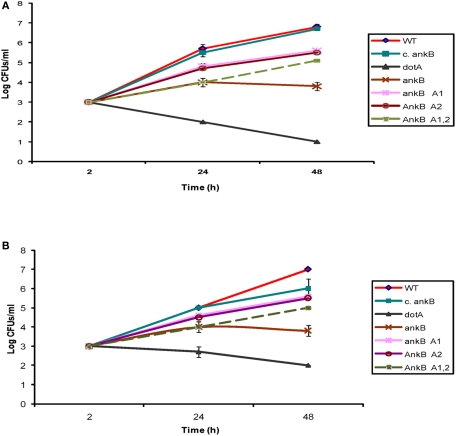
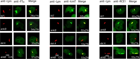
The Dot/Icm-translocated Ankyrin B (AnkB) F-box effector of Legionella pneumophila is essential for intra-vacuolar proliferation and functions as a platform for the docking of polyubiquitinated proteins to the Legionella-containing vacuole (LCV) within macrophages and ameba. Here we show that ectopically expressed AnkB in Dictyostelium discoideum is targeted to the plasma membrane where it recruits polyubiquitinated proteins and it trans-rescues the intracellular growth defect of the ankB null mutant, which has never been demonstrated for any effector in ameba. Using co-immunoprecipitation and bimolecular fluorescence complementation we show specific interaction of Skp1 of D. discoideum with the F-box domain of AnkB, which has never been demonstrated in ameba. We show that anchoring of AnkB to the cytosolic face of the LCV membrane in D. discoideum is mediated by the host farnesylation of the C-terminal eukaryotic CaaX motif of AnkB and is independent of the F-box and the two ANK domains, which has never been demonstrated in ameba. Importantly, the three host farnesylation enzymes farnesyl transferase, RCE-1, and isoprenyl cysteine carboxyl methyl transferase of D. discoideum are recruited to the LCV in a Dot/Icm-dependent manner, which has never been demonstrated in ameba. We conclude that the polyubiquitination and farnesylation enzymatic machineries of D. discoideum are recruited to the LCV in a Dot/Icm-dependent manner and the AnkB effector exploits the two evolutionarily conserved eukaryotic machineries to proliferate within ameba, similar to mammalian cells. We propose that L. pneumophila has acquired ankB through inter-kingdom horizontal gene transfer from primitive eukaryotes, which facilitated proliferation of L. pneumophila within human cells and the emergence of Legionnaires' disease.

Keywords: SCF1; Skp1; dot/Icm; farnesyl; prenylation.


Figures

Figure 1
Figure 1
Biological function of AnkB as platforms for the recruitment of polyubiquitinated proteins to the plasma membrane of AnkB-transfected D. discoideum. D. discoideum was transfected with the FLAG-AnkB, or FLAG-AnkB169C/A, FLAG-ΔAnkB1,2, FLAG-ΔF-box, or AnkB9L10P/AA. Localization of FLAG-AnkB fusion proteins was examined by confocal laser scanning microscopy. Cells were labeled with anti-AnkB antibodies (green) and anti-polyubiquitin antibodies (red). The data are representatives of three independent experiments.
Figure 2
Figure 2
Trans-rescue of the ankB null mutant for intra-ameba growth defect in AnkB-transfected D. discoideum. Representative confocal microscopy images of D. discoideum to determine the formation of replicative LCVs. (A) Untransfected and (B) Transfected D. discoideum with FLAG-AnkB were infected with either the WT strain or the ΔankB mutant for 1 h and examined at 2 and 12 h post-infection. Cells were labeled with anti-Lpn antibodies (blue) and anti-AnkB antibodies (green). Rescue was determined by the observations of replicative vacuoles for the ankB mutant. The data are representatives of three independent experiments.
Figure 3
Figure 3
In vivo interaction of AnkB with Skp1 of D. discoideum. (A) Representative confocal images of co-transfected D. discoideum with constructs expressing fusions of AnkB-YN and Skp1-YC or AnkB-ΔF-box-YN and Skp1-YC. The data are representatives of three independent experiments. (B) D. discoideum were infected with L. pneumophila strains for 2 h. Skp1 was immunoprecipitated from semi-purified LCVs using anti-AnkB antibodies and then analyzed by immunoblotting with anti-AnkB antibodies followed by anti-Skp1 antibodies. The experiments were performed twice and representative examples are shown.
Figure 4
Figure 4
Ankyrin B is modified by the host cell farnesylation machinery. D. discoideum were infected with the L. pneumophila strains. The infection was performed for 1 h and the cells were examined at 2 h post-infection. The AnkB proteins were immunoprecipitated from semi-purified LCVs using anti-AnkB antibodies and then analyzed by immunoblotting with anti-AnkB and by anti-farnesyl antibodies. The data are representatives of independent experiments.
Figure 5
Figure 5
Farnesylation by D. discoideum anchors AnkB to the LCV membrane. The infection was performed for 1 h and the cells were lysed at 2 h post-infection. The LCVs were isolated from untreated or FTI-277-treated D. discoideum. The LCVs were labeled with (A) anti-AnkB antibodies prior to permeabilization (green). After permeabilization, the LCVs were labeled with anti-L. pneumophila (Lpn, red). (B). The LCVs were permeabilized then labeled with anti-AnkB and anti-L. pneumophila antibodies. Samples were analyzed by confocal microscopy and analyses were based on examination of 100 LCVs from different coverslips from triplicate samples. The data are representatives of three independent experiments.
Figure 6
Figure 6
The two ANK domains are dispensable for anchoring AnkB to the cytosolic face of the LCV membrane. The infection was performed for 1 h and the cells were lysed at 2 h post-infection to purify the LCVs. Semi-purified LCVs were analyzed by confocal microscopy. Representative confocal microscopy images that show location of AnkB at the cytosolic face of LCVs. (A) The LCVs were probed with anti-SidC prior to permeabilization of membranes (green). After permeabilization of membranes, the LCVs were stained with DAPI to visualize L. pneumophila (Lpn blue). (B) The LCVs were permeabilized then labeled with anti-SidC and DAPI stain. (C,D) Integrity of the membrane of semi-purified LCVs from D. discoideum was verified by (C) labeling with anti-AnkB antibodies (green) prior to permeabilization of the LCVs. After permeabilization, the LCVs were labeled with anti-Lpn antibodies (blue) within the LCVs. (D) The LCVs were permeabilized followed by labeling with anti-AnkB and anti-Lpn antibodies (blue). Quantification is shown in the merged panels, where the numbers represent the percentage +SD of LCVs that showed localization of AnkB to the cytosolic face of the LCV membrane. Quantitation was based on analyses of 100 LCVs from different coverslips. The data are representatives of three independent experiments.
Figure 7
Figure 7
The two ANK domains of AnkB are essential for the biological function of AnkB in intracellular growth of L. pneumophila in D. discoideum. (A) D. discoideum or (B) A. polyphaga were infected with the WT strain, the ankB mutant, or the ankB mutant harboring one of the mutant alleles ankBΔA1, ankBΔA2, or ankBΔA1A2. The ankB mutant complemented with native WT ankB (c.ankB) and the dotA were used as controls. The infection was carried out for 1 h using an MOI of 10 followed by treatment with gentamicin for 1 h. The infected monolayers were lysed at different time points and plated onto agar plates for colony enumeration. The results are representative of three independent experiments performed in duplicate.
Figure 8
Figure 8
Dot/Icm-dependent recruitment of the three farnesylation enzymes FTα, RCE-1, and Icmt to the LCV within D. discoideum. D. discoideum were infected with various strains for 1 h. At 2 h after infection, the cells were labeled with anti-Lpn antibodies (red) and anti-FTα, anti-RCE-1, or anti-IcmT antibodies (green) and analyzed by confocal microscopy, and analyses were based on examination of 100 LCVs from different coverslips from triplicate samples. The data are representatives of three independent experiments.

Similar articles

Cited by

References

    1. Al-Khodor S., Price C. T., Habyarimana F., Kalia A., Abu Kwaik Y. (2008). A Dot/Icm-translocated ankyrin protein of Legionella pneumophila is required for intracellular proliferation within human macrophages and protozoa. Mol. Microbiol. 70, 908–923 - PMC - PubMed
    1. Al-Khodor S., Price C. T., Kalia A., Abu Kwaik Y. (2010). Functional diversity of ankyrin repeats in microbial proteins. Trends Microbiol. 18, 132–13910.1016/j.tim.2009.11.004 - DOI - PMC - PubMed
    1. Amer A. O. (2010). Modulation of caspases and their non-apoptotic functions by Legionella pneumophila. Cell. Microbiol. 12, 140–147 - PubMed
    1. Berger K. H., Isberg R. R. (1993). Two distinct defects in intracellular growth complemented by a single genetic locus in Legionella pneumophila. Mol. Microbiol. 7, 7–19 - PubMed
    1. Bergo M. O., Leung G. K., Ambroziak P., Otto J. C., Casey P. J., Young S. G. (2000). Targeted inactivation of the isoprenylcysteine carboxyl methyltransferase gene causes mislocalization of K-Ras in mammalian cells. J. Biol. Chem. 275, 17605–17610 - PubMed

LinkOut - more resources