. 2011 Jun 1:2:118.
doi: 10.3389/fmicb.2011.00118. eCollection 2011.

Genetic and Functional Diversity of Pseudomonas aeruginosa Lipopolysaccharide

Affiliations

Genetic and Functional Diversity of Pseudomonas aeruginosa Lipopolysaccharide

Joseph S Lam et al. Front Microbiol. .

Abstract

Lipopolysccharide (LPS) is an integral component of the Pseudomonas aeruginosa cell envelope, occupying the outer leaflet of the outer membrane in this Gram-negative opportunistic pathogen. It is important for bacterium-host interactions and has been shown to be a major virulence factor for this organism. Structurally, P. aeruginosa LPS is composed of three domains, namely, lipid A, core oligosaccharide, and the distal O antigen (O-Ag). Most P. aeruginosa strains produce two distinct forms of O-Ag, one a homopolymer of D-rhamnose that is a common polysaccharide antigen (CPA, formerly termed A band), and the other a heteropolymer of three to five distinct (and often unique dideoxy) sugars in its repeat units, known as O-specific antigen (OSA, formerly termed B band). Compositional differences in the O units among the OSA from different strains form the basis of the International Antigenic Typing Scheme for classification via serotyping of different strains of P. aeruginosa. The focus of this review is to provide state-of-the-art knowledge on the genetic and resultant functional diversity of LPS produced by P. aeruginosa. The underlying factors contributing to this diversity will be thoroughly discussed and presented in the context of its contributions to host-pathogen interactions and the control/prevention of infection.

Keywords: bacteriophage; biosynthesis; lipopolysaccharide; motility; nucleotide sugars; seroconversion; serotyping; virulence.


Figures
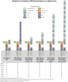
Figure 1
Figure 1
Representations of the heterogeneity/diversity of the LPS glycoforms present on the surface of a single P. aeruginosa cell. Genetic defects in members of various assembly pathway genes and their resultant changes to the variety of LPS glycoforms present have been indicated, with “✓” or “✗” representing their presence or absence, respectively. Substitutions of various lipid A and core OS sugars with phosphate groups and L-Ala have been indicated with yellow circles and red diamonds, respectively. The OSA polymer from serotype O5 has been displayed as a representative polymer.
Figure 2
Figure 2
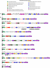
Organization of the genes within OSA biosynthesis clusters. (A) OSA biosynthesis cluster of serotype O5 adapted from Burrows et al. (1996). The gene cluster is located on the complementary strand; genes which match the PFAM designation are colored accordingly. Genes not involved in OSA biosynthesis are depicted in gray including a large insertion sequence (IS). (B) Adapted from Raymond et al. (2002), the OSA biosynthesis gene clusters were organized into 11 groups based on sequence conservation. Genes were designated using the PFAM database; specific protein families which occur a minimum of three times throughout all 20 OSA biosynthesis clusters are represented by a specific color. A red outline depicts an ORF with potential transmembrane-spanning domains. Previously identified genes are labeled above the respected cluster if present within the serotype. Insertion sequences (IS) present within genes are depicted by a secondary gray box.
Figure 3
Figure 3
Proposed CPA gene clusters of (A) P. aeruginosa PAO1, (B) PA14, (C) LESB58, (E) PA7, and (D) P. fluorescens pfO-1. The organism-specific identifier for numbered genes of unknown function is indicated in brackets beside each organism.
Figure 4
Figure 4
Structures of uncapped and capped glycoforms. The structures of the uncapped core oligosaccharide (A), and capped core oligosaccharide (B) are depicted. All the sugars shown have α configuration unless otherwise indicated. Asterisks depict variable substitutions, acetylation sites are shown since they have not been precisely identified. Ala, alanine; Cm, carbamoyl; Etn, ethanolamine; GalN, 2-amino-2-deoxy-galactose (galactosamine); Glc, glucose; Hep, L-glycero-d-manno-heptose; Kdo, 3-deoxy-d-manno-oct-2-ulosonic acid; Rha, rhamnose. Adapted from King et al. (2009).
Figure 5
Figure 5
Different lipid A forms of P. aeruginosa and the proposed biosynthesis and modification pathway.
Figure 6
Figure 6
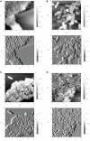
mages obtained from Atomic Force Microscopy analyses of P. aeruginosa cells collected in contact mode in air. (A) Strain PAO1; (B) migA mutant; (C) wapR mutant; (D) rmlC mutant. Height images (top panels) are shown with enclosed areas for roughness calculations. Deflection images (bottom panels) reveal more details in cell morphology. The straight lines in (A) are steps in the mica substratum. Reproduced from Lau et al. (2009b), with permission from Copyright Clearance Centre.
Figure 7
Figure 7
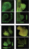
Images from confocal laser scanning microscopy analyses of P. aeruginosa cells that illustrate the changes in biofilm structure resulting from truncation in the LPS core in mutant strains as comparing to the wildtype bacteria. Average projections (top panels) and midpoint cross sections (bottom panels) of representative microcolonies of (A) wildtype strain PAO1, (B) migA mutant, (C) wapR mutant, and (D) rmlC mutant are shown. Reproduced from Lau et al. (2009b), with permission from Copyright Clearance Centre.

References

    1. Abeyrathne P. D., Daniels C., Poon K. K., Matewish M. J., Lam J. S. (2005). Functional characterization of WaaL, a ligase associated with linking O-antigen polysaccharide to the core of Pseudomonas aeruginosa lipopolysaccharide. J. Bacteriol. 187, 3002–301210.1128/JB.187.9.3002-3012.2005 - DOI - PMC - PubMed
    1. Abeyrathne P. D., Lam J. S. (2007). WaaL of Pseudomonas aeruginosa utilizes ATP in in vitro ligation of O antigen onto lipid A-core. Mol. Microbiol. 65, 1345–1359 - PubMed
    1. Anderson P. (2010). Emerging therapies in cystic fibrosis. Ther. Adv. Respir. Dis. 4, 177–185 - PubMed
    1. Arsenault T. L., Hughes D. W., Maclean D. B., Szarek W. A., Kropinski A. M. B., Lam J. S. (1991). Structural studies on the polysaccharide portion of ‘A-band’ lipopolysaccharide from a mutant (AK14O1) of Pseudomonas aeruginosa PAO1. Can. J. Chem. 69, 1273–1280
    1. Augustin D. K., Song Y., Baek M. S., Sawa Y., Singh G., Taylor B., Rubio-Mills A., Flanagan J. L., Wiener-Kronish J. P., Lynch S. V. (2007). Presence or absence of lipopolysaccharide O antigens affects type III secretion by Pseudomonas aeruginosa. J. Bacteriol. 189, 2203–220910.1128/JB.01839-06 - DOI - PMC - PubMed

LinkOut - more resources