. 2011:2011:657460.
doi: 10.4061/2011/657460. Epub 2011 Jun 1.

Trypanosoma cruzi Coexpressing Ornithine Decarboxylase and Green Fluorescence Proteins as a Tool to Study the Role of Polyamines in Chagas Disease Pathology

Affiliations

Trypanosoma cruzi Coexpressing Ornithine Decarboxylase and Green Fluorescence Proteins as a Tool to Study the Role of Polyamines in Chagas Disease Pathology

Jeremías José Barclay et al. Enzyme Res. 2011.

Abstract

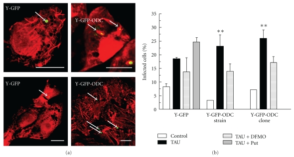
Polyamines are essential for Trypanosoma cruzi, the causative agent of Chagas disease. As T. cruzi behaves as a natural auxotrophic organism, it relies on host polyamines biosynthesis. In this paper we obtained a double-transfected T. cruzi parasite that expresses the green fluorescent protein (GFP) and a heterologous ornithine decarboxylase (ODC), used itself as a novel selectable marker. These autotrophic and fluorescent parasites were characterized; the ODC presented an apparent Km for ornithine of 0.51 ± 0.16 mM and an estimated V(max) value of 476.2 nmoles/h/mg of protein. These expressing ODC parasites showed higher metacyclogenesis capacity than the auxotrophic counterpart, supporting the idea that polyamines are engaged in this process. This double-transfected T. cruzi parasite results in a powerful tool-easy to follow by its fluorescence-to study the role of polyamines in Chagas disease pathology and in related processes such as parasite survival, invasion, proliferation, metacyclogenesis, and tissue spreading.


Figures

Figure 1
Figure 1
Growth kinetics of Y-GFP and Y-GFP-ODC clone cultured at different conditions. Cultures carried out under the indicated conditions were diluted to 10–20 × 106 cells/mL when the stationary phase was reached. (a) Y-GFP cultured in rich media BHT (- -- -), Diamond (—■—), and in Diamond supplemented with 2 mg/mL glucose (- -△- -). (b) Y-GFP cultured in the semisynthetic medium SDM79 without () or with (- -- -) 1 mM putrescine. (c) Y-GFP-ODC clone cultured in SDM79 (——) or in SDM79 supplemented with 5 mM α-difluoromethylornithine (DFMO) (- -- -). All cultures were continuously maintained in presence of 250 μg/mL G418.
Figure 2
Figure 2
Fluorescence microscopy of Y-GFP (a) and Y-GFP-ODC strain (b) and the selected Y-GFP-ODC clone epimastigotes (c) (1000x).
Figure 3
Figure 3
Ornithine decarboxylase (ODC) specific activity in Y-GFP and Y-GFP-ODC cell extracts. (a) ODC specific activity was measured in cell extracts obtained at different times after transfection; (—■—) Y-GFP strain; (- -- -) Y-GFP-ODC strain. (b) ODC activity in extracts of exponentially growing cultures at 60 days after transfection and clone selection. All values are the average of at least three experiments.
Figure 4
Figure 4
ODC activity characterization. (a) Plot of Lineweaver-Burk. ODC activity was assayed in vitro at different concentrations of the substrate ornithine to estimate apparent Km and Vmax. (b) The Y-GFP-ODC clone was cultured in absence or presence of 5 mM α-difluoromethylornithine (DFMO) for 24 hs, then cells were harvested, washed and ODC activity was measured. (c) ODC activity was carried out in the presence of DFMO at different concentrations. Values in B and C are given as percentages of maximal activity. All other details as indicated in Materials and Methods. These values are the average of two independent experiments.
Figure 5
Figure 5
Metacyclogenesis of Y-GFP and Y-GFP-ODC strain and clone. After metacyclogenesis protocol (see details in Materials and Methods), the total mixed suspensions of E and MT from each condition (Control, TAU, or TAU + DFMO) were used to infect Vero cells for 24 h followed by detection of the host cell actin cytoskeleton as indicated under Materials and Methods. (a) Confocal images showing Vero cells infected for 24 h with T. cruzi Y-GFP and Y-GFP-ODC clone obtained from TAU medium. (b) Percentage of infected Vero cells with T. cruzi Y-GFP, T. cruzi Y-GFP-ODC strain, and T. cruzi Y-GFP-ODC clone at indicated conditions. Data represent the mean ± SEM of at least two independent experiments (number of counted cells 100). Significantly different from control: **P < .01, Bars: 10 μm.

References

    1. Heby O, Persson L, Rentala M. Targeting the polyamine biosynthetic enzymes: a promising approach to therapy of African sleeping sickness, Chagas’ disease, and leishmaniasis. Amino Acids. 2007;33(2):359–366. - PubMed
    1. Schofield CJ, Maudlin I. Trypanosomiasis control. International Journal for Parasitology. 2001;31(5-6):615–620. - PubMed
    1. Andrade LO, Andrews NW. Opinion: the Trypanosoma cruzi—host-cell interplay: location, invasion, retention. Nature Reviews Microbiology. 2005;3(10):819–823. - PubMed
    1. Tyler KM, Engman DM. The life cycle of Trypanosoma cruzi revisited. International Journal for Parasitology. 2001;31(5-6):472–481. - PubMed
    1. Kohl L, Robinson D, Bastin P. Novel roles for the flagellum in cell morphogenesis and cytokinesis of trypanosomes. The EMBO Journal. 2003;22(20):5336–5346. - PMC - PubMed