. 2011;6(6):e20975.
doi: 10.1371/journal.pone.0020975. Epub 2011 Jun 8.

Preconditioning involves selective mitophagy mediated by Parkin and p62/SQSTM1

Affiliations

Preconditioning involves selective mitophagy mediated by Parkin and p62/SQSTM1

Chengqun Huang et al. PLoS One. 2011.

Abstract

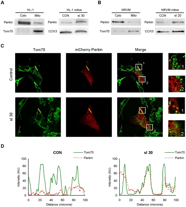
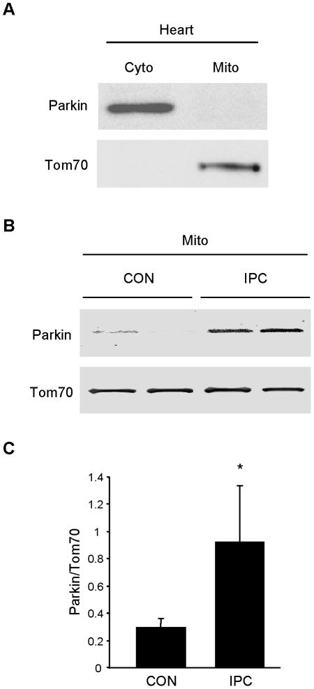
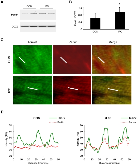
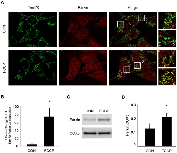
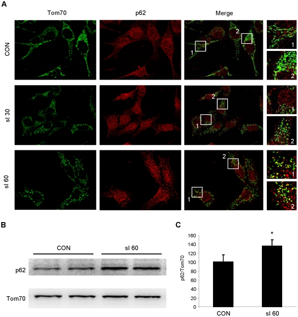
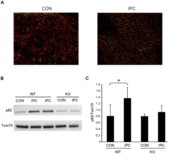
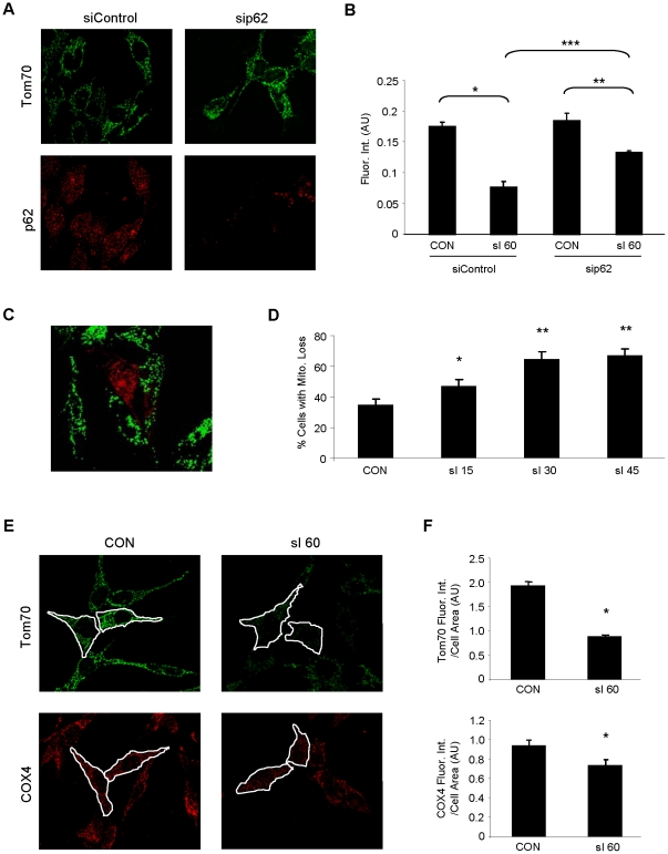
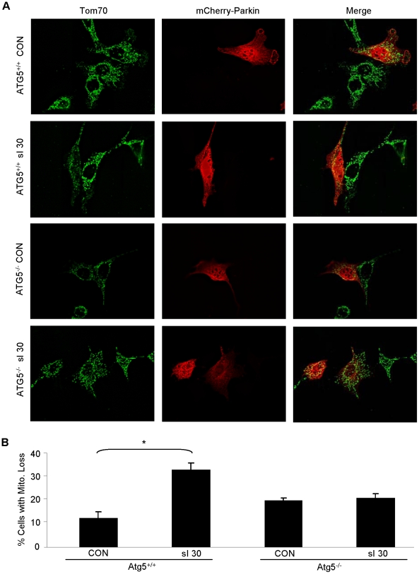
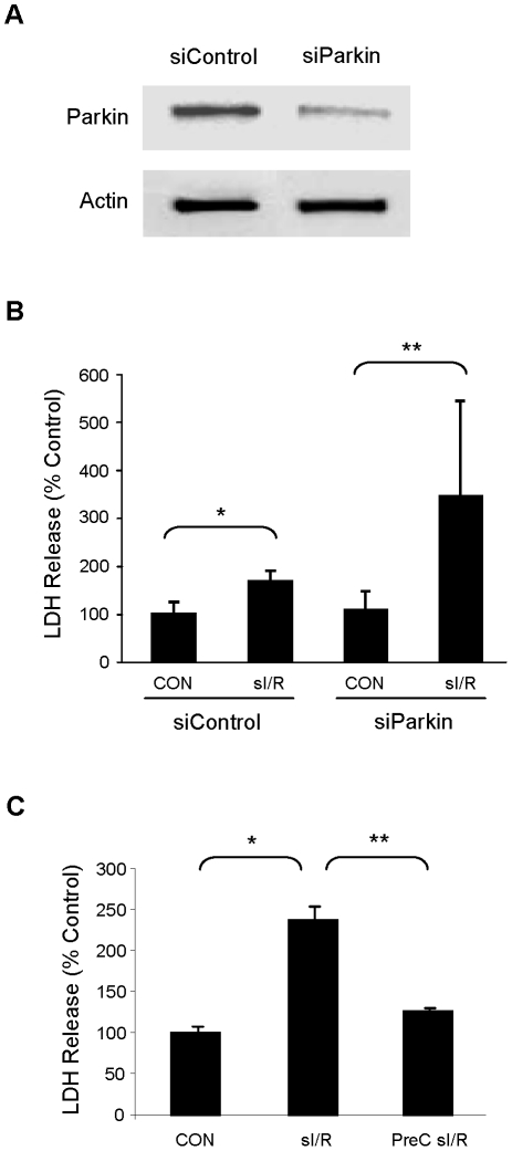
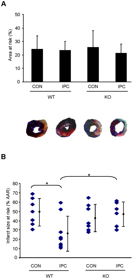
Autophagy-dependent mitochondrial turnover in response to cellular stress is necessary for maintaining cellular homeostasis. However, the mechanisms that govern the selective targeting of damaged mitochondria are poorly understood. Parkin, an E3 ubiquitin ligase, has been shown to be essential for the selective clearance of damaged mitochondria. Parkin is expressed in the heart, yet its function has not been investigated in the context of cardioprotection. We previously reported that autophagy is required for cardioprotection by ischemic preconditioning (IPC). In the present study, we used simulated ischemia (sI) in vitro and IPC of hearts to investigate the role of Parkin in mediating cardioprotection ex vivo and in vivo. In HL-1 cells, sI induced Parkin translocation to mitochondria and mitochondrial elimination. IPC induced Parkin translocation to mitochondria in Langendorff-perfused rat hearts and in vivo in mice subjected to regional IPC. Mitochondrial depolarization with an uncoupling agent similarly induced Parkin translocation to mitochondria in cells and Langendorff-perfused rat hearts. Mitochondrial loss was blunted in Atg5-deficient cells, revealing the requirement for autophagy in mitochondrial elimination. Consistent with previous reports indicating a role for p62/SQSTM1 in mitophagy, we found that depletion of p62 attenuated mitophagy and exacerbated cell death in HL-1 cardiomyocytes subjected to sI. While wild type mice showed p62 translocation to mitochondria and an increase in ubiquitination, Parkin knockout mice exhibited attenuated IPC-induced p62 translocation to the mitochondria. Importantly, ablation of Parkin in mice abolished the cardioprotective effects of IPC. These results reveal for the first time the crucial role of Parkin and mitophagy in cardioprotection.


Conflict of interest statement

Competing Interests: The authors have declared that no competing interests exist.

Figures

Figure 1
Figure 1. Parkin redistributes to mitochondria in cardiomyocytes subjected to simulated ischemia (sI).
A. HL-1 cells were subjected to simulated ischemia (ischemia-mimetic buffer and hypoxia) for the indicated time, then fractionated to yield cytosol and heavy membranes (mitochondria-enriched fraction). Right-hand panel shows Parkin in the heavy membrane fraction under basal conditions (CON) and after 30 min of simulated ischemia (sI 30). B. Neonatal rat ventricular cardiomyocytes were subjected to simulated ischemia, then fractionated to yield crude cytosol and heavy membrane fractions. Right-hand panel shows Parkin in the heavy membrane fraction under resting conditions (CON) and after 20 min sI. C. HL-1 cells transfected with mCherry-Parkin (red) were subjected to 30 min sI, then fixed and immunolabeled with mitochondrial marker Tom70 (green). Yellow line indicates the segment used for pseudo-line scan analysis. Boxes indicate regions enlarged at right. Images are representative of 5 independent replicates. D. Pseudo-line-scan tracing indicates distribution of mitochondria (Tom70, solid green line) and mCherry-Parkin (dotted red line).
Figure 2
Figure 2. Parkin translocates in response to IPC in rat hearts.
A. Under resting conditions, Parkin is restricted to cytosol in the heart. B. Langendorff-perfused rat hearts were subjected to continuous perfusion (CON) or 3 cycles of 5 min global no-flow ischemia alternating with 5 min reperfusion (IPC). Immediately after the third 5 min reperfusion, hearts were homogenized and fractionated to yield crude cytosol and heavy membrane fractions which were probed for Parkin. C. Quantification of Parkin in mitochondrial fractions from CON and IPC hearts is shown (*p<0.05, n = 4).
Figure 3
Figure 3. IPC triggers Parkin translocation to mitochondria in vivo.
A. Mice were subjected to IPC consisting of 3 cycles of 5 min coronary artery ligation alternating with 5 min reperfusion. Heavy membrane fractions prepared from heart were probed for Parkin. Cytochrome oxidase subunit III (COX3) was used to normalize mitochondrial loading. B. Quantification of Parkin translocation to mitochondria is shown (*p<0.05, n = 5). C. Mice were subjected to 3 cycles of IPC in vivo. Cryosections of hearts were examined to visualize Parkin translocation to mitochondria by immunolabeling with antibodies to Tom70 (green) and Parkin (red). White line indicates segment used for pseudo-line scan analysis. Representative images from the risk zone of sham-operated (CON) and preconditioned (IPC) hearts are shown. D. Pseudo-line scan analysis of mitochondria (Tom70, solid green line) and endogenous Parkin (dotted red line) in cryosections from control (CON) and preconditioned (IPC) mouse hearts demonstrates translocation of Parkin to the mitochondria after IPC.
Figure 4
Figure 4. FCCP induces Parkin translocation to mitochondria in HL-1 cardiomyocytes and mitochondrial fractions in Langendorff-perfused rat hearts.
A. HL-1 cardiomyocytes were treated with the mitochondrial uncoupler FCCP (10 µM) or vehicle (ethanol, CON) for 1 hour, then fixed and immunolabeled for Tom70 (green) and Parkin (red). Boxes outline fields that were enlarged (at right) to show details of mitochondrial structure and colocalization. B. Cells were then scored for significant colocalization. Over 100 cells from sequential fields were assessed for each group (*p<0.03, n = 4). C. Isolated rat hearts were subjected to continuous perfusion with 100 nM FCCP or vehicle (ethanol, CON) for 5 minutes after stabilization with KHB. Mitochondrial fractions were probed for Parkin. D. Quantification of Parkin in mitochondrial fractions from CON and FCCP treated hearts (*p<0.05, n = 4).
Figure 5
Figure 5. p62 translocates to mitochondria in HL-1 cells subjected to simulated ischemia (sI).
A. HL-1 cells were subjected to sI for 30 or 60 min, then fixed and immunolabeled for Tom70 (green) and p62 (red). Boxes outline fields that were enlarged (at right) to show details of mitochondrial structure and colocalization. B. p62 Western blot of mitochondrial fractions from HL-1 cardiomyocytes subjected to 60 min sI. C. Quantification of Western blots is shown (Student's T-test: *p<0.02, n = 3).
Figure 6
Figure 6. Translocation of p62 to mitochondria is induced by IPC.
A. Cryosectioned hearts from WT mice sham operated (CON) or subject to IPC were immunolabeled for p62 and Tom70. Colocalization was assessed via PDM value images using Image J software where high colocalization scores are shown in green. B. WT and Parkin−/− (KO) mice were subjected to sham surgery (CON) or IPC, and mitochondria were probed for p62 and normalized to Tom70. C. Quantification of p62 translocation is shown (*p<0.05, n = 5).
Figure 7
Figure 7. Depletion of p62 attenuates sI-induced mitochondrial loss.
A. HL-1 cells were treated with scrambled siRNA (siControl) or siRNA corresponding to p62 (sip62) and labeled with antibodies to Tom70 (green) and p62 (red). B. HL-1 cells treated with siRNA were subjected to sI for 60 min and mitochondrial mass was quantified by fluorescence intensity of the mitochondrial marker Tom70. Over 100 cells were assessed for each group and the experiment was performed 3 times. Error bars represent SEM of the 3 experiments (ANOVA: *p<0.001, **p<0.05, ***p<0.005). C. Shown is a representative deconvolved image of HL-1 cells transfected with mCherry-Parkin (red) and subjected to sI for 30 min and probed for Tom70 (green). D. Mitochondrial content in mCherry-Parkin-transfected cells was assessed by fluorescence intensity of Tom70, and the percentage of cells showing substantial mitochondrial loss (similar to the cell depicted here in the center of the field) was scored for non-ischemic cells (CON) and time points of 15, 30 and 45 min of simulated ischemia. A minimum of 100 transfected cells were scored for each time point (ANOVA: *p<0.01, **p<0.001 versus CON, n = 4). Error bars represent SEM of the 4 experiments. E. Mitochondrial content per cell was assessed in HL-1 cardiomyocytes subjected to 60 min of sI. The total amount of green (Tom70, outer mitochondrial membrane marker), and red (COX4, inner mitochondrial membrane marker) fluorescence intensity per unit area within each cell were measured (ImageJ). F. For each condition, 100 cells were scored in sequential fields (*p<0.01, n = 4).
Figure 8
Figure 8. Autophagy is necessary for selective mitochondrial loss mediated by Parkin after simulated ischemia.
A. MEFs from ATG5+/+ and ATG5−/− mice were transfected with mCherry-Parkin (red) and then subjected to 30 min sI. After fixation, mitochondria were visualized with antibodies to Tom70 (green). Representative deconvolved images are shown. B. Mitochondrial depletion was quantified in a minimum of 50 mCherry-Parkin expressing cells per condition (*p<0.01, n = 4).
Figure 9
Figure 9. Parkin mediates cytoprotection in HL-1 cells subjected to simulated ischemia and reperfusion (sI/R).
A. HL-1 cells were treated with siRNA for Parkin or control siRNA and lysates were probed for Parkin protein. Equal protein loading is shown by blotting for actin. B. Cells treated with siRNA were subjected to sI/R (60 min simulated ischemia and 60 min reperfusion) and cell death was determined by measuring LDH release into the culture medium (Student's T-Test: *p<0.05, **p<0.001, n = 6). C. HL-1 cells were incubated in normal media for 60 min, subjected to sI/R, or preconditioned (PreC) with 30 min sI and 60 min recovery in normal media before being subjected to sI/R. Cell culture supernatants during the final 60 min reperfusion were assayed for LDH release to determine cell death. (*p<0.005, and **p<0.01, n = 3).
Figure 10
Figure 10. Parkin is required for IPC.
A. WT and Parkin−/− (KO) mice were subjected to sham treatment (CON) or IPC, followed by 20 min ischemia and 22 hr reperfusion. Area at risk was determined by Evan's blue injection after re-occlusion and showed no difference between groups. B. Infarct size was determined by TTC staining and reported as % of the area at risk. IPC reduced infarct size in WT mice but not in Parkin KO mice (*p<0.05, n = 6). Representative heart slices are shown.

References

    1. Willis MS, Patterson C. Hold me tight: Role of the heat shock protein family of chaperones in cardiac disease. Circulation. 2010;122:1740–1751. - PMC - PubMed
    1. Goldberg AL. Protein degradation and protection against misfolded or damaged proteins. Nature. 2003;426:895–899. - PubMed
    1. Geisler S, Holmstrom KM, Skujat D, Fiesel FC, Rothfuss OC, et al. PINK1/Parkin-mediated mitophagy is dependent on VDAC1 and p62/SQSTM1. Nat Cell Biol. 2010;12:119–131. - PubMed
    1. Mizushima N, Levine B, Cuervo AM, Klionsky DJ. Autophagy fights disease through cellular self-digestion. Nature. 2008;451:1069–1075. - PMC - PubMed
    1. Vives-Bauza C, Zhou C, Huang Y, Cui M, de Vries RL, et al. PINK1-dependent recruitment of Parkin to mitochondria in mitophagy. Proc Natl Acad Sci U S A. 2010;107:378–383. - PMC - PubMed

Publication types

MeSH terms

LinkOut - more resources