. 2010 Nov 11:1:123.
doi: 10.3389/fmicb.2010.00123. eCollection 2010.

Molecular Detection of Legionella: Moving on From mip

Affiliations

Molecular Detection of Legionella: Moving on From mip

Stacey F Y Yong et al. Front Microbiol. .

Abstract

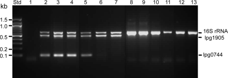
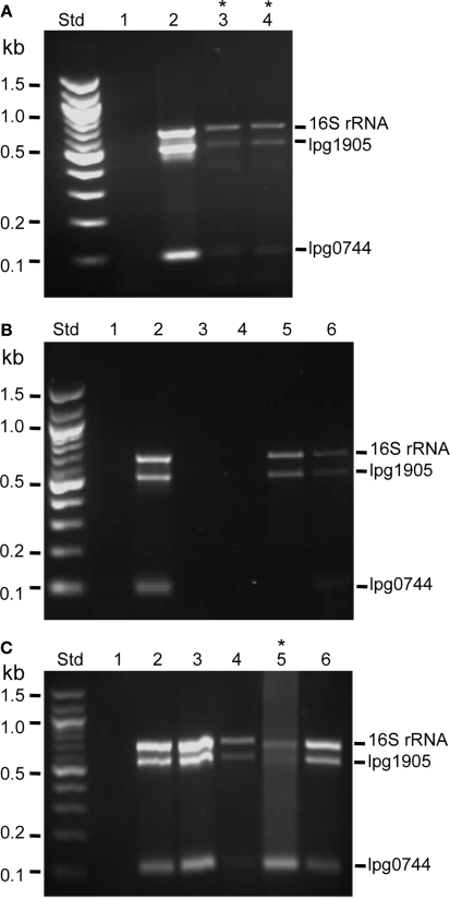
The detection of Legionella pneumophila in environmental and clinical samples is frequently performed by PCR amplification of the mip and/or 16S rRNA genes. Combined with DNA sequencing, these two genetic loci can be used to distinguish different species of Legionella and identify L. pneumophila. However, the recent Legionella genome sequences have opened up hundreds of possibilities for the development of new molecular targets for detection and diagnosis. Ongoing comparative genomics has the potential to fine tune the identification of Legionella species and serogroups by combining specific and general genetic targets. For example, the coincident detection of LPS biosynthesis genes and virulence genes may allow the differentiation of both pathogen and serogroup without the need for nucleotide sequencing. We tested this idea using data derived from a previous genomic subtractive hybridization we performed between L. pneumophila serogroup 1 and L. micdadei. Although not yet formally tested, these targets serve as an example of how comparative genomics has the potential to improve the scope and accuracy of Legionella molecular detection if embraced by laboratories undertaking Legionella surveillance.

Keywords: Legionella; genomics; molecular testing; virulence.


Figures

Figure 1
Figure 1
Representative gel showing simultaneous detection of lpg1905, lpg0774, and 16SrRNA by multiplex PCR. Std, 100 base pair marker (Promega); Lane 1, no DNA control; Lane 2, L. pneumophila serogroup 1 strain 02/41; Lane 3, L. pneumophila serogroup 1 strain B6; Lane 4, L. pneumophila serogroup 1 strain CS1; Lane 5, L. pneumophila serogroup 1 strain 02/41; Lane 6, L. pneumophila serogroup 2–14 strain C11(1); Lane 7, L. pneumophila serogroup 2–14 strain C4(1); Lane 8, L. gormanii strain C9; Lane 9, L. anisa strain L041; Lane 10, L. gormanii 03/69; 11, L. longbeachae A4C5; 12, L. longbeachae ATCC33462; 13, L. longbeachae Atlanta 5. For lpg0774 (Gene Bank: AY688227) the upstream primer started at base 46: 5′-TGCTAACAACCACTATCCCAAA-3′ and downstream primer started at base 202: 5′-GTTTCAATAAAAGCGTGCTCCT-3′. The upstream primer of lpg1905 (Gene Bank: NC_002942) started at base 328: 5′-TTGCCTAAAACTCACCACAGAA-3′ and downstream primer started at base 857: 5′-5′ATGCCGCCCAAAATATACC-3′. The 16S rRNA primers included in the triplex PCR to identify the genus Legionella have been described previously (Miyamoto et al., 1997). Triplex PCR was performed using 20 ng of template DNA in a 25 μL PCR reaction mix containing 1 × Green GoTaq® Flexi Buffer (Promega), 2 mM MgCl2, 200 μM dNTP, 0.5 μM of each primer and 1 U GoTaq® DNA polymerase. The optimized triplex PCR condition was performed in MyCycler™ (BIORAD) at initial denaturation of 95°C for 4 min followed by 35 cycles of 95°C for 1 min, 57.5°C for 1 min, and 72°C for 1 min with a final extension at 72°C for 5 min. The amplified products were then analyzed by DNA gel electrophoresis.
Figure 2
Figure 2
Detection of lpg1905, lpg0774, and 16SrRNA in environmental and clinical samples by multiplex PCR. (A) Detection of Legionella spp. in cooling towers at an office building. Std, 100 base pair marker (Promega); Lane 1, negative control (no DNA); Lane 2, L. pneumophila serogroup 1 strain 02/41; Lane 3, Cooling tower 1; Lane 4, Cooling tower 2. (B) Detection of Legionella spp. in cooling tower and shower head water samples collected from a hotel. Std, 100 base pair marker (Promega); Lane 1, negative control (no DNA); Lane 2, L. pneumophila serogroup 1 strain 02/41; Lane 3, Cooling tower surface water; Lane 4, Cooling tower sediment; Lane 5, Shower head 915; Lane 6, Shower head 1617. (C) Detection of Legionella spp. in patient samples. Std, 100 base pair marker (Promega); Lane 1, negative control (no DNA); Lane 2, L. pneumophila serogroup 1 strain 02/41; Lane 3, Sputum spiked with L. pneumophila serogroup 1 strain 02/41; Lane 4, Patient 1 (male, 22 years old), sputum sample; Lane 5, Patient 2 (male, 70 years old), bronchial wash; Lane 6, Patient 3 (male, 56 years old), sputum sample. In the above examples, 500 ml of cooling tower water was collected by immersing a sterilized 1000 ml bottle approximately 10 cm below water surface. To collect a showerhead sample, hot water was turned on for 5 min prior to collection of 50 ml of sample. Water samples were pressure filtered through a 0.45 μm cellulose nitrate membrane (Millipore), and eluted with 5 ml of sterile phosphate buffered saline (PBS pH 7.2). Following centrifugation (4,000 rpm, 20 min) the resulting sediment was resuspended in 2 ml sterile distilled water. Total DNA was extracted from 200 μl of the sediment suspension using the QIAamp DNA Mini Kit (Qaigen, Germany). A further 1 ml of sediment suspension was treated with 9 ml of HCl–KCl pH 2.2 (0.1 M Tris HCl, 0.1 M KCl) for 20 min then cultured onto BCYE agar containing GVPC selective supplement (Oxoid). 100 ml of the non-acid treated sediment suspension was also diluted 10-fold and cultured on BCYE-GVPC media. Total DNA was extracted from patient sputum and bronchial washes using QIAamp DNA blood mini kit (Qiagen). To liquefy viscose and sticky sputum samples, 40 μl of freshly prepared sputasol (0.75% [wt/vol]) (Oxoid) was added into 200 μl of sputum sample and the mixture was incubated at 37°C for 30 min (Bencini et al., 2007). The remaining 200 μl sputum or bronchial wash was cultured on BCYE media containing BMPA selective supplement (Oxoid). * indicates samples that were also positive for L. pneumophila by bacteriological culture.

Similar articles

Cited by

References

    1. Aurell H., Etienne J., Forey F., Reyrolle M., Girardo P., Farge P., Decludt B., Campese C., Vandenesch F., Jarraud S. (2003). Legionella pneumophila serogroup 1 strain Paris: endemic distribution throughout France. J. Clin. Microbiol. 41, 3320–332210.1128/JCM.41.7.3320-3322.2003 - DOI - PMC - PubMed
    1. Bencini M. A., van den Brule A. J., Claas E. C., Hermans M. H., Melchers W. J., Noordhoek G. T., Salimans M. M., Schirm J., Vink C., van der Zee A., Jansen R. (2007). Multicenter comparison of molecular methods for detection of Legionella spp. in sputum samples. J. Clin. Microbiol. 45, 3390–339210.1128/JCM.00505-07 - DOI - PMC - PubMed
    1. Benin A. L., Benson R. F., Arnold K. E., Fiore A. E., Cook P. G., Williams L. K., Fields B., Besser R. E. (2002). An outbreak of travel-associated Legionnaires disease and Pontiac fever: the need for enhanced surveillance of travel-associated legionellosis in the United States. J. Infect. Dis. 185, 237–24310.1086/338060 - DOI - PubMed
    1. Cazalet C., Gomez-Valero L., Rusniok C., Lomma M., Dervins-Ravault D., Newton H. J., Sansom F. M., Jarraud S., Zidane N., Ma L., Bouchier C., Etienne J., Hartland E. L., Buchrieser C. (2010). Analysis of the Legionella longbeachae genome and transcriptome uncovers unique strategies to cause Legionnaires’ disease. PLoS Genet. 6, e1000851 10.1371/journal.pgen.1000851 - DOI - PMC - PubMed
    1. Cazalet C., Jarraud S., Ghavi-Helm Y., Kunst F., Glaser P., Etienne J., Buchrieser C. (2008). Multigenome analysis identifies a worldwide distributed epidemic Legionella pneumophila clone that emerged within a highly diverse species. Genome Res. 18, 431–44110.1101/gr.7229808 - DOI - PMC - PubMed

LinkOut - more resources