. 2010 Nov 11:1:125.
doi: 10.3389/fmicb.2010.00125. eCollection 2010.

Role of phospholipases in fungal fitness, pathogenicity, and drug development - lessons from cryptococcus neoformans

Affiliations

Role of phospholipases in fungal fitness, pathogenicity, and drug development - lessons from cryptococcus neoformans

Julianne Teresa Djordjevic. Front Microbiol. .

Abstract

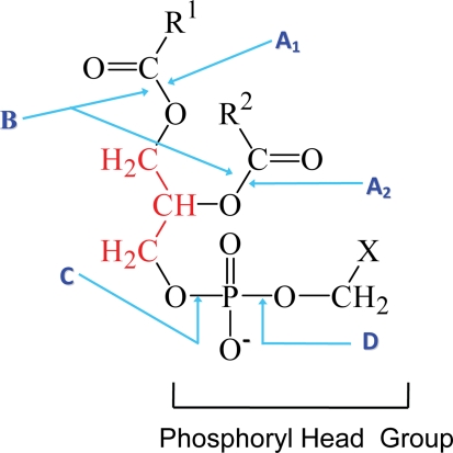
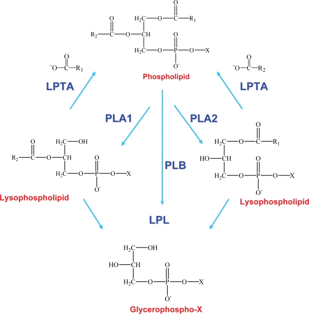
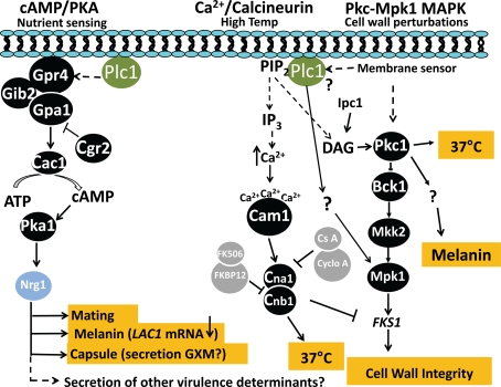
Many pathogenic microbes, including many fungi, produce phospholipases which facilitate survival of the pathogen in vivo, invasion and dissemination throughout the host, expression of virulence traits and evasion of host immune defense mechanisms. These phospholipases are either secreted or produced intracellularly and act by physically disrupting host membranes, and/or by affecting fungal cell signaling and production of immunomodulatory effectors. Many of the secreted phospholipases acquire a glycosylphosphatidylinositol sorting motif to facilitate membrane and/or cell wall association and secretion. This review focuses primarily on the role of two members of the phospholipase enzyme family, phospholipase B (Plb) and phosphatidylinositol (PI)-specific phospholipase C (PI-C/Plc), in fungal pathogenesis and in particular, what has been learnt about their function from studies performed in the model pathogenic yeast, Cryptococcus neoformans. These studies have revealed how Plb has adapted to become an important part of the virulence repertoire of pathogenic fungi and how its secretion is regulated. They have also provided valuable insight into how the intracellular enzyme, Plc1, contributes to fungal fitness and pathogenicity - via a putative role in signal transduction pathways that regulate the production of stress-protecting pigments, polysaccharide capsule, cell wall integrity, and adaptation to growth at host temperature. Finally, this review will address the role fungal phospholipases have played in the development of a new class of antifungal drugs, which mimic their phospholipid substrates.

Keywords: Cryptococcus neoformans; PI-PLC/Plc; drug development; pathogenicity; phosphatidylinositol-specific phospholipase C; phospholipase B; secretion; signaling.


Figures

Figure 1
Figure 1
The phospholipid cleavage sites of phospholipase enzymes. The phospholipid glycerol backbone is shown in red. Phospholipase A1 (A1) cleaves a fatty acyl ester at the sn1 position while phospholipase A2 (A2) removes the fatty acid at the sn2 position to yield a lysophospholipid and a fatty acid. Phospholipase B (B) removes both acyl chains from the sn1 and sn2 positions on the glycerol backbone. The phosphodiester bond in the head group proximal to the glycerol backbone is cleaved by phospholipase C (C) to yield diacylglycerol and the phosphoryl head group. PI-PLC enzymes perform this cleavage when X is inositol, which may, or may not, be linked to phosphates or a sugar chain composed predominantly of mannosyl sugars in the case of a GPI anchor. Cleavage by phospholipase D (D) at the phosphodiester bond proximal to X, yields phosphatidic acid and X, which can be either ethanolamine, serine, inositol, or choline. Adapted from the PhD thesis of Dr Rosemary A. Siafakas.
Figure 2
Figure 2
Enzymatic activities of phospholipase B enzymes. Phospholipase B (PLB) activity is a combination of PLA1 and PLA2 activities (or PLA and LPL activity) and generates glycerophospho compounds. Lysophospholipid, a short-lived intermediate, is also a substrate for lysophospholipase (LPL) and lysophospholipase-transacylase (LPTA) activities, which rapidly hydrolyse or re-esterify lysophospholipid, respectively. R1 and R2 represent fatty acid chains in the sn1 and sn2 position of the phospholipid, respectively. X refers to the head group (e.g., serine, ethanolamine, choline, or inositol). Adapted from the PhD thesis of Dr Rosemary A. Siafakas.
Figure 3
Figure 3
Proposed role of CnPlc1 in pathogenicity-related signaling in C. neoformans. 1. Cyclic AMP (cAMP)/Protein kinase A (Pka) signaling pathway. A change in extracellular nutrient levels trigger activation of the integral membrane-associated G-protein coupled receptor, GPR4, causing its associated Gα subunit (Gpa1) to activate adenylyl cyclase (Cac1), resulting in production of cAMP. cAMP binds to regulatory subunits (Pkr1) of the Pka complex (not shown) to release an active form of the catalytic subunit (Pka1), which phosphorylates the transcription factor, Nrg1. A GTPase-activating protein (Crg2) negatively regulates Gpa1. Gib2, Gβ subunit. Based on studies in S. cerevisiae and our own published and preliminary data in C. neoformans demonstrating the requirement for CnPlc1 in melanin production (via LAC1 transcription), capsule production and secretion of virulence determinants (Chayakulkeeree et al., 2008), CnPlc1 may be linked to cAMP signaling possibly by regulating the activation of Gpa1 as indicated by the dashed line. Lac1, Laccase 1; GXM, glucuronic acid-xylose-mannan. 2. Ca2+/calcineurin signaling pathway. This pathway primarily regulates high temperature growth in C. neoformans, but not in S. cerevisiae. High temperatures may trigger CnPlc1 to hydrolyse PIP2, producing IP3, which potentially raises intracellular Ca2+ (see dashed line) by regulating Ca2+ pumps/channels in the plasma membrane and ER. Cytosolic Ca2+ is sensed by calmodulin (Cam1), which activates calmodulin kinases (CaMKs) (not shown) and the calcineurin complex (Cna1 and Cnb1) to support growth at 37°C. Cyclosporine A (CsA) and FK506, bind cyclophilin A and FKBP12, respectively, to inhibit calcineurin; PIP2, phosphatidylinositol 4,5-bisphosphate. Calcineurin is also essential for hyphal elongation during mating and monokaryotic fruiting (not shown). 3. Pkc1/Mpk1 MAPK signaling pathway. This pathway conveys cell wall perturbation signals detected by unknown membrane sensors, to the nucleus. CnPlc1 is essential for activation of Mpk1 in response to cell wall perturbation in a process that may, or may not be, mediated by activation of Pkc1 via the CnPlc1 hydrolysis product, DAG (see dashed line). Activated Pkc1 phosphorylates MAPKKK (Bck1) and subsequent phosphorylations of MAPKK (Mkk2) by Bck1 and MAPK (Mpk1) by Mkk2 to upregulate transcription of FKS1, which encodes β-1,3-glucan synthase. Expression of FKS1 is also negatively regulated by calcineurin. DAG produced by the sphingolipid synthetic pathway enzyme inositolphosphoceramide 1 synthase (Ipc1), and possibly by CnPlc1, also activates CnPkc1 to regulate melanin production independently of Mpk1 activation (Heung et al., 2005). Pkc1 is also required for host temperature growth (Gerik et al., 2008).

Similar articles

Cited by

References

    1. Aimanianda V., Clavaud C., Simenel C., Fontaine T., Delepierre M., Latge J. P. (2009). Cell wall beta-(1,6)-glucan of Saccharomyces cerevisiae: structural characterization and in situ synthesis. J. Biol. Chem. 284, 13401–1341210.1074/jbc.M807667200 - DOI - PMC - PubMed
    1. Alspaugh J. A., Perfect J. R., Heitman J. (1997). Cryptococcus neoformans mating and virulence are regulated by the G-protein alpha subunit GPA1 and cAMP. Genes Dev. 11, 3206–3217 - PMC - PubMed
    1. Alspaugh J. A., Pukkila-Worley R., Harashima T., Cavallo L. M., Funnell D., Cox G. M., Perfect J. R., Kronstad J. W., Heitman J. (2002). Adenylyl cyclase functions downstream of the Galpha protein Gpa1 and controls mating and pathogenicity of Cryptococcus neoformans. Eukaryotic Cell 1, 75–8410.1101/gad.11.23.3206 - DOI - PMC - PubMed
    1. Alvarez M., Burn T., Luo Y., Pirofski L. A., Casadevall A. (2009). The outcome of Cryptococcus neoformans intracellular pathogenesis in human monocytes. BMC Microbiol. 9, 51.10.1128/EC.1.1.75-84.2002 - DOI - PMC - PubMed
    1. Alvarez M., Casadevall A. (2006). Phagosome extrusion and host-cell survival after Cryptococcus neoformans phagocytosis by macrophages. Curr. Biol. 16, 2161–216510.1186/1471-2180-9-51 - DOI - PubMed