. 2011 Jun 3:5:38.
doi: 10.3389/fnsys.2011.00038. eCollection 2011.

Loss of specificity in Basal Ganglia related movement disorders

Affiliations

Loss of specificity in Basal Ganglia related movement disorders

Maya Bronfeld et al. Front Syst Neurosci. .

Abstract

The basal ganglia (BG) are a group of interconnected nuclei which play a pivotal part in limbic, associative, and motor functions. This role is mirrored by the wide range of motor and behavioral abnormalities directly resulting from dysfunction of the BG. Studies of normal behavior have found that BG neurons tend to phasically modulate their activity in relation to different behavioral events. In the normal BG, this modulation is highly specific, with each neuron related only to a small subset of behavioral events depending on specific combinations of movement parameters and context. In many pathological conditions involving BG dysfunction and motor abnormalities, this neuronal specificity is lost. Loss of specificity (LOS) manifests in neuronal activity related to a larger spectrum of events and consequently a large overlap of movement-related activation patterns between different neurons. We review the existing evidence for LOS in BG-related movement disorders, the possible neural mechanisms underlying LOS, its effects on frequently used measures of neuronal activity and its relation to theoretical models of the BG. The prevalence of LOS in a many BG-related disorders suggests that neuronal specificity may represent a key feature of normal information processing in the BG system. Thus, the concept of neuronal specificity may underlie a unifying conceptual framework for the BG role in normal and abnormal motor control.

Keywords: Parkinson's disease; Tourette syndrome; basal ganglia; dyskinesia; dystonia; information encoding; movement.


Figures

Figure 1
Figure 1
Neuronal specificity. Peri-event time histograms and raster plots of the activity of a single neuron around the onset-time of different movements (simulated data), illustrating the concept of neuronal specificity. (A) Specific encoding, the neuron displays rate changes only in relation to movements of a single joint. (B) Non-specific encoding, the neuron displays similar rate modulations related to movements of multiple joints.
Figure 2
Figure 2
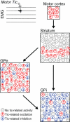
Non-specific tic-related neuronal activity. Schematic illustration depicting the relative frequency and spatial distribution of neuronal activity patterns in the cortico-basal ganglia system related to motor tics confined to a single muscle group (Bronfeld et al., 2011). In the striatum, tic-related activity was confined to the somatotopical territory of the tic location, but almost all projection neurons recorded from that territory displayed non-specific tic-related bursts of activity. In the pallidum, tic-related activity was detected in a very large and spatially diffuse subpopulation of neurons. Tic-related excitations and inhibitions were the predominant responses in GPe and GPi neurons, respectively.
Figure 3
Figure 3
Neural mechanisms underlying neuronal specificity. Schematic illustrations of intra-nucleus (A) and inter-nuclei (B) mechanisms underlying neuronal specificity. Red and blue filled circles represent excited and inhibited neurons, respectively. (A) In the striatum, inhibitory projections from GABAergic interneurons and collaterals from neighboring projection neurons (MSNs) enable selective activation of some MSNs while inhibiting others, in response to common excitatory cortical inputs (Based on: Tepper et al., 2004). (B) In the GPi, the combined effects of spatially focused inhibitory projections from the striatum, and spatially diffused excitatory projections from the subthalamic nucleus enable selective movement-related excitation/inhibition activity patterns (Based on: Mink, 1996).
Figure 4
Figure 4
Effect of loss of specificity on properties of neuronal activity. (A) Illustration of firing rate fluctuations of multiple neurons, depicting rate modulations related to different movements, in a neuronal population for which rate increases are the predominant pattern of movement-related activity. Left: Specific encoding, each neuron modulates its firing rate in response to a single type of movement. Right: Loss of specificity (LOS); each neuron displays similar movement-related rate modulations in response to a larger range of movements. (B) Following LOS, the fraction of movement-related neurons is increased. (C) The number of movements eliciting rate modulations in each neuron increases, resulting in an increased effect of movements on the overall average firing rate of each neuron (i). For a population in which most neurons have a dominant non-specific response pattern (such as firing rate increase), LOS has an effect on the overall average firing rate of the population (ii). (D) Non-specific encoding of movements generates correlations between pairs of neurons (top panel). These correlations represent only the common rate modulations attributed to the commonly encoded movements, and following subtraction of these effects (middle panel, the shift predictor), the normalized correlation function shows no correlation between the neurons (bottom panel).
Figure 5
Figure 5
Theoretical models of the basal-ganglia. In all panels blue and red arrows indicate inhibitory and excitatory effects, respectively. (A) Schematic representation of the BG circuitry, according to the “box and arrow” model. (Based on: Albin et al., ; DeLong, 1990). (B) Illustration of BG “action selection process”: integration of excitatory and inhibitory pathways through the BG lead to focused excitation and diffused “surround” inhibition of thalamic neurons, thereby selecting the desired motor pattern while inhibiting potentially competing motor patterns (Based on: Mink, 1996). (C) Structure of the cortico-BG neural network according to the “reinforcement driven dimensionality reduction” model. Information from multiple cortical regions is integrated by lateral inhibitory connections within the striatum and feed-forward projections from the striatum to the GPi. A reward signal is provided via dopaminergic (DA) projections, and used to modulate the relative significance of different input dimensions (Based on: Bar-Gad et al., 2000).

Similar articles

Cited by

References

    1. Agid Y. (1991). Parkinson's disease: pathophysiology. Lancet 337, 1321–1324 - PubMed
    1. Albin R. L., Mink J. W. (2006). Recent advances in Tourette syndrome research. Trends Neurosci. 29, 175–182 - PubMed
    1. Albin R. L., Young A. B., Penney J. B. (1989). The functional anatomy of basal ganglia disorders. Trends Neurosci. 12, 366–375 - PubMed
    1. Alexander G. E., Crutcher M. D. (1990a). Functional architecture of basal ganglia circuits: neural substrates of parallel processing. Trends Neurosci. 13, 266–271 - PubMed
    1. Alexander G. E., Crutcher M. D. (1990b). Neural representations of the target (goal) of visually guided arm movements in three motor areas of the monkey. J. Neurophysiol. 64, 164–178 - PubMed

LinkOut - more resources