

In vivo three-dimensional segmental analysis of adolescent idiopathic scoliosis
- PMID: 21688001
- PMCID: PMC3175875
- DOI: 10.1007/s00586-011-1869-4
In vivo three-dimensional segmental analysis of adolescent idiopathic scoliosis
Abstract
Introduction: An accurate assessment of three-dimensional (3D) intervertebral deviation is crucial to the better surgical correction of adolescent idiopathic scoliosis (AIS). However, a precise 3D study of intervertebral deviation has not been previously reported.
Objective: The purpose of the present study is to evaluate the intervertebral coronal inclination, axial rotation and sagittal angulation of AIS using 3D bone models and a local coordinate system.
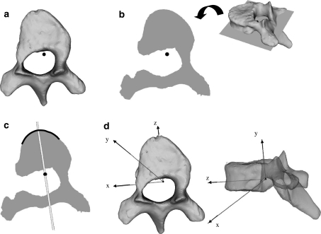
Materials and methods: 3D bone models of the thoracic and lumbar spine of ten AIS patients were constructed using computed tomography. The local coordinate axis was determined semi-automatically for each vertebra. By using these local coordinates, the intervertebral deviation angles were calculated in the coronal, axial and sagittal planes and projected to subjacent local coordinates.
Result: The intervertebral deformity in the coronal plane was larger near the apical region and smaller near the junctional region. Conversely, the intervertebral rotation in the axial plane was smaller near the apical region, and larger near the junctional region. Concerning the sagittal plane deformity, the constant tendency was not recognized.
Conclusion: Using a local coordinate system for the vertebra of AIS, we measured the 3D intervertebral coronal, axial and sagittal deviation of the thoracolumbar spine and found that the change in the intervertebral inclination angle in the coronal plane increased toward the apical region and decreased toward the junctional region, and that the converse tendency was noted for the axial intervertebral rotational angle. This analysis provides an improved 3D guide for the surgical correction of AIS.
Figures


References
-
- Asghar J, Samdani AF, Pahys JM, D’andrea LP, Guille JT, Clements DH, Betz RR. Computed tomography evaluation of rotation correction in adolescent idiopathic scoliosis: a comparison of an all pedicle screw construct versus a hook–rod system. Spine. 2009;34:804–807. doi: 10.1097/BRS.0b013e3181996c1b. - DOI - PubMed
-
- Benameur S, Mignotte M, Parent S, Labelle H, Skalli W, Guise J. 3D biplanar statistical reconstruction of scoliotic vertebrae. Stud Health Technol Inform. 2002;91:281–285. - PubMed
-
- Cruickshank JL, Koike M, Dickson RA (1989) Curve patterns in idiopathic scoliosis. A clinical and radiographic study. J Bone Joint Surg [Br] 71:259–263 - PubMed
-
- Dubousset J. Three-dimensional analysis of the scoliotic deformity. In: Weinstein SL, editor. The pediatric Spine: Principles and Practices. New York: Raven Press; 1994. pp. 479–496.
MeSH terms
LinkOut - more resources
Full Text Sources
Medical
