. 2011 Jul 25;50(31):7052-5.
doi: 10.1002/anie.201101266. Epub 2011 Jun 17.

Membrane anchored immunostimulatory oligonucleotides for in vivo cell modification and localized immunotherapy

Affiliations

Membrane anchored immunostimulatory oligonucleotides for in vivo cell modification and localized immunotherapy

Haipeng Liu et al. Angew Chem Int Ed Engl. .
No abstract available


Figures

Figure 1
Figure 1
In vitro screening for optimal ODN conjugate structures. a) Schematic illustration of lipophilic-ODN insertion into cell membranes. b) Flow cytometric evaluation of membrane anchoring efficiency by different liphophilic modifications, left: flow cytometry histograms. black: untreated B16F10 cells, red: C18 single chain lipid ODN, blue: diacyllipid-PEG-ODN, purple: cholesterol-ODN and green: diacyllipid-ODN. Right: relative insertion efficiencies of each ODN conjugate based on the mean fluorescence intensity. c) Molecular structure of diacyllipid ODN. d) Confocal image of diacyllipid ODN-modified B16 cells. e) After 2 h of culture at 37 °C, a partial internalization of ODNs can be observed. Scale bar: 50 μm.
Figure 2
Figure 2
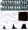
In vivo cell modification by lipo-ODNs. a,b) In vivo kinetics of fluorescence decay of rhodamine-conjugated lipo-ODN (a) and non-lipidated ODN (b). The upper sites on mice were subcutaneous injections into healthy tissue; the lower sites were intratumoral injections. c) Quantification of total fluorescence over time from IVIS whole-animal imaging of injection sites. d, e) Representative confocal image of tumor cells recovered from an intratumoral injection site (d) and flow cytometric analysis of recovered tumor cells (e) 3 h after injection. Scale bar: 50 μm.
Figure 3
Figure 3
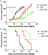
Therapeutic effects of lipid modified CpG ODN. a) Time course analysis of tumor growth (n=10) after treated with two injections of either lipo-GpC, CpG or lipo-CpG. The differences for the treatment of lipo-CpG versus CpG were statistically significant (P<0.004, paired t-test). b) Kaplan–Meier survival curve (with log-rank test) after treated with ODN probes, tumor-bearing mice treated with lipo-CpG have a prolonged survival compared with CpG group (P<0.01).

References

    1. Bubeník J. J. Cancer Res. Clin. Oncol. 1990;116:1–7. - PubMed
    1. Gimbel MI, Delman MIKA, Zager JS. Cancer Control. 2008;15:225–232. - PubMed
    1. Riker AI, Radfar S, Liu S, Wang Y, Khong HT. Expert Opin. Biol. Ther. 2007;7:345–358. - PubMed
    1. Vollmer J, Krieg AM. Adv. Drug Delivery Rev. 2009;61:195–204. - PubMed
    1. Wooldridge JE, Weiner GJ. Curr. Opin. Oncol. 2003;15:440–445. - PubMed

Publication types