Case Reports
. 2011 Jun 20:8:314.
doi: 10.1186/1743-422X-8-314.

Capacity building permitting comprehensive monitoring of a severe case of Lassa hemorrhagic fever in Sierra Leone with a positive outcome: case report

Affiliations
Case Reports

Capacity building permitting comprehensive monitoring of a severe case of Lassa hemorrhagic fever in Sierra Leone with a positive outcome: case report

Jessica N Grove et al. Virol J. .

Abstract

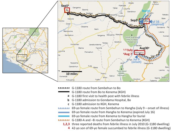
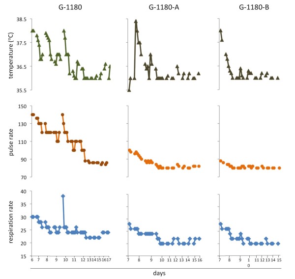
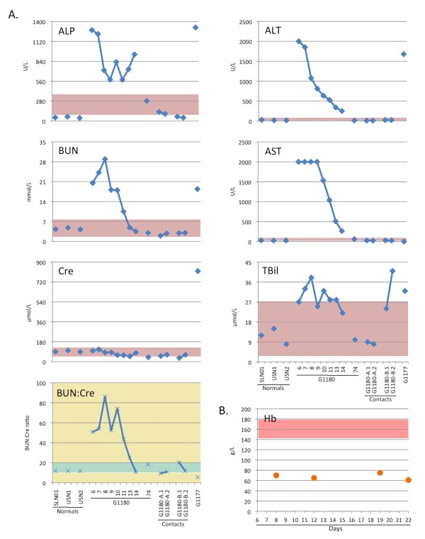
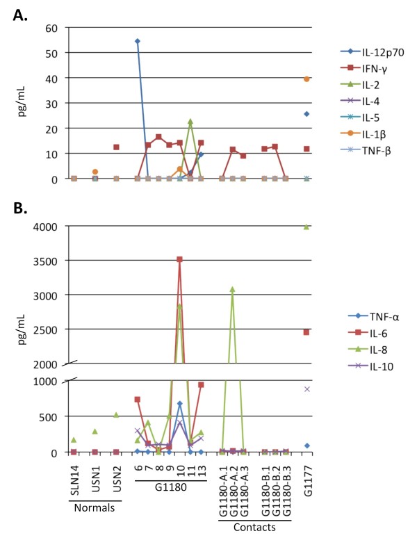
Lassa fever is a neglected tropical disease with a significant impact on the health care system of endemic West African nations. To date, case reports of Lassa fever have focused on laboratory characterisation of serological, biochemical and molecular aspects of the disease imported by infected individuals from Western Africa to the United States, Canada, Europe, Japan and Israel. Our report presents the first comprehensive real time diagnosis and characterization of a severe, hemorrhagic Lassa fever case in a Sierra Leonean individual admitted to the Kenema Government Hospital Lassa Fever Ward. Fever, malaise, unresponsiveness to anti-malarial and antibiotic drugs, followed by worsening symptoms and onset of haemorrhaging prompted medical officials to suspect Lassa fever. A recombinant Lassa virus protein based diagnostic was employed in diagnosing Lassa fever upon admission. This patient experienced a severe case of Lassa hemorrhagic fever with dysregulation of overall homeostasis, significant liver and renal system involvement, the interplay of pro- and anti-inflammatory cytokines during the course of hospitalization and an eventual successful outcome. These studies provide new insights into the pathophysiology and management of this viral illness and outline the improved infrastructure, research and real-time diagnostic capabilities within LASV endemic areas.


Figures

Figure 1
Figure 1
Map of Sierra Leone and expanded view of Eastern Province, with relevant localities and routes travelled by patients in the current case report. The Eastern Province of Sierra Leone is shown in the inset map with location of Sembehun, where all suspected Lassa fever cases in the current report originated, and the three localities where patients travelled to and from: Bo, Kenema, and Hangha. The inbound routes travelled by each patient are indicated in dotted lines, and outbound ones in solid lines. Three deaths from febrile illness in July were reported to the Outreach team during a local investigation in Sembehun following admission of G-1180 to KGH LFW. The travel history of a 69 year old female and her daughter from Sembehun to Hangha, and subsequently to Kenema, where she succumbed to a febrile illness without final diagnosis are highlighted. The 42 year old son of the 69 year old female reportedly succumbed to a similar febrile illness with hemorrhage on August 7 in Sembehun, without a final diagnosis. G-1180 was first transported to Ghondama clinic in Bo on September 1, after being referred by the local health post in Sembehun on August 30th with fever and general malaise. The patient was subsequently transported to KGH LFW on September 2nd after Lassa fever was suspected based on clinical presentation.
Figure 2
Figure 2
Vital signs for G-1180 and contacts -A and -B during hospitalization at KGHLFW. Core temperature (°C) [▲], pulse [●], and respiratory rates (per minute) [◆] were measured at regular intervals, usually every 4 hours at the onset, and every 12 hours at later times, throughout the hospitalization period for each patient. Data points were plotted individually and the normal ranges for each parameter are indicated by a line (temperature) or boxes (pulse and respiratory rates). A. Patient G-1180. B. G-1180 contacts -A and B.
Figure 3
Figure 3
A comprehensive metabolic panel was obtained daily by Piccolo analysis. Fourteen metabolic indicators were measured in the serum of G-1180 daily after admission, through day 14 (with the exception of day 12), using a Piccolo comprehensive metabolic panel disk array (see additional file 1 figure S1 for Na+, Cl-, Ca2+, K+, TCO2, Alb and TP levels).A Two samples from G-1180 contacts -A and -B were also analyzed, along with Sierra Leonean and U.S. normal controls. Values were plotted alongside normal ranges for each metabolite, for reference (rose boxes). G-1180 presented with elevated TBil, and extremely high BUN:Cre ratios. Surprisingly, levels of Cre remained within normals levels throughout, in contrast to other report cases of Lassa hemorrhagic fever. The most dramatically elevated metabolic indicators were ALP, ALT, and AST, all indicative of severe liver implication in this case of Lassa fever. Patient G-1177, who succumbed to Lassa fever, presented with elevated TBil, but low BUN:Cre, mainly due to extremely high Cre levels. This patient also had highly elevated levels of ALP and ALT, but normal levels of AST. Metabolic indicators were assayed in normal Sierra Leonean and U.S. normals, along with two samples each from G-1180 contacts -A and -B. B Haemoglobin levels (Hb) were measured in G-1180 on days 8, 12, 19, and 22 post- onset of disease. Low levels of Hb prompted blood transfusions on days 9 and 14.
Figure 4
Figure 4
Serum cytokine levels analyzed by multiplex Flow Cytometry. Serum cytokine levels were analyzed with a BenderMed Systems Human 11-Plex Inflammatory Cytokine kit, and an Accuri C6 Flow Cytometer equipped with dual laser detection. Data was processed and quantitated with Flow Cytomic Pro software, and plotted on linear scales. G-1180 presented with elevated levels of IL-12p70, IL-6, and IL-10, all of which decreased by the following day. On day 10 post onset of disease a significant but transient spike in IL-1β, IL-6, IL-8, TNF-α, and IL-10 occurred, that rapidly decreased overnight, mostly to background levels. The only notable cytokine fluctuation in G-1180 contacts occurred in -A.2 with a dramatic spike in IL-8 that decreased to background levels within 2 days (-A.3). The single point analysis for G-1177 revealed high levels of IL-1β, IL12p70, IL-6, and IL-10.
Figure 5
Figure 5
Rapid diagnosis of acute Lassa fever virus infection by Lateral Flow Immunoassay (LFI) in patient G-1180 and contacts -A and -B. LFI tests contain a murine monoclonal antibody capture and an heterologous gold-conjugated antibody for the detection of LASV nucleoprotein (NP) in the serum of suspected Lassa fever patients. Twenty-five μL of serum were applied to the sample well in an LFI module and chased with 3 drops (~ 100 μL) of optimally formulated buffer. After 10 minutes the results were recorded photographically. A representative normal serum sample analysis from a Sierra Leonean donor (S.L. N01) is shown for comparison. Only the control line developed with this serum sample. Conversely, sera from G-1180 generated a detectable precipitate in the test line, indicative of LASV NP antigen. The LFI platform detected NP antigen from days 6 - 8. Days 9 and 10 show no detectable antigen in this format. Sera from G-1180 contacts -A and -B did not produce a detectable signal on LFI, despite detection of antigen by a sensitive NP ELISA (figure 4A).
Figure 6
Figure 6
ELISA detection of LASV NP antigen and virus-specific immunoglobulins M and G in normal donors, G-1180, G-1180 contacts, and in two additional patient and contact sera (G-1177 and G-1177-A). An antigen capture ELISA designed with affinity-purified goat polyclonal antibody reagents was used to detect LASV NP in patient sera (A). LASV NP antigen was not detected in normal sera from Sierra Leone and U.S. origin, or in most G-1180 contacts. The level of LASV NP antigen [◆, blue] in G-1180 dropped significantly during the first 3 days of ribavirin administration (quantitative levels indicated), to nearly undetectable levels by day 11. Conversely, antigen levels in G-1180-A and –B did not drop over the course of three days of ribavirin treatment. G-1180-F registered a significant level of antigen but did not seek treatment. G-1177 succumbed to acute Lassa fever, with very high levels of viral antigen detected before expiry. LASV-specific IgM (B) [▲, red] and IgG (C) [●, green] were detected in a recombinant ELISA plate format, coated with NP, GP1, and GP2, or with individually coated proteins. Most Sierra Leonean sera showed significant levels of IgM, IgG, or both, whereas U.S. normals did not. IgM and IgG levels in G-1180 rose throughout the course of the illness, and remained high on day 74 post onset of infection (black arrow). For G-1180 data are also plotted with IgM (▬ NP, ■ GP1, ◆ GP2, green) and IgG (▬ NP, ■ GP1, ◆ GP2, red) responses to individual LASV proteins. The IgM and IgG responses were directed primarily against NP, with a low IgG titer to GP1detected on day 74. Data are plotted as mean ±SD, N=2.
Figure 7
Figure 7
Composite analysis of all parameters evaluated for G-1180 in the course of these studies. Data for core temperature, respiration and pulse rates, LASV antigen, virus-specific IgM and IgG, metabolites, and cytokines were aligned for facile comparison of daily profiles. The single profile on day 10, which registered an increase in core body temperature to febrile levels, dramatic increase in respiratory and pulse rates early in the day, accompanied by elevated levels of pro- and anti-inflammatory cytokines is noteworthy, and is boxed in white.

References

    1. Buckley SM, Casals J. Lassa fever, a new virus disease of man from West Africa. 3. Isolation and characterization of the virus. Am J Trop Med Hyg. 1970;19(4):680–91. - PubMed
    1. Birmingham K, Kenyon G. Lassa fever is unheralded problem in West Africa. Nat Med. 2001;7(8):878.. - PubMed
    1. Fisher-Hoch SP, McCormick JB. Lassa fever vaccine: A review. Expert Rev Vaccines. 2004;3:103–111. - PubMed
    1. McCormick JB, King IJ, WEbb, Chomczynski PA, Johnson KM, O'Sullivan R, Smith ES, Trippel S, Tong TC, Sacchi N. A case-control study of the clinical diagnosis and course of Lassa fever. J. Infect Dis. 1987;155(3):445–455. doi: 10.1093/infdis/155.3.445. - DOI - PubMed
    1. McCormick JB. Epidemiology and control of Lassa fever. Current Topics in Microbiol and Immunol. 1987;134:69–78. - PubMed

Publication types

LinkOut - more resources