. 2011 Jun 23;70(6):1085-99.
doi: 10.1016/j.neuron.2011.04.025.

Isoform-specific dephosphorylation of dynamin1 by calcineurin couples neurotrophin receptor endocytosis to axonal growth

Affiliations

Isoform-specific dephosphorylation of dynamin1 by calcineurin couples neurotrophin receptor endocytosis to axonal growth

Daniel Bodmer et al. Neuron. .

Abstract

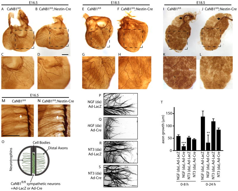
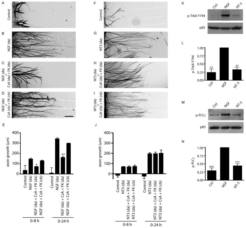
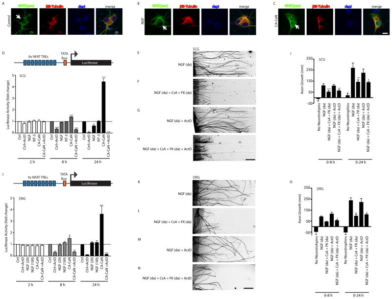
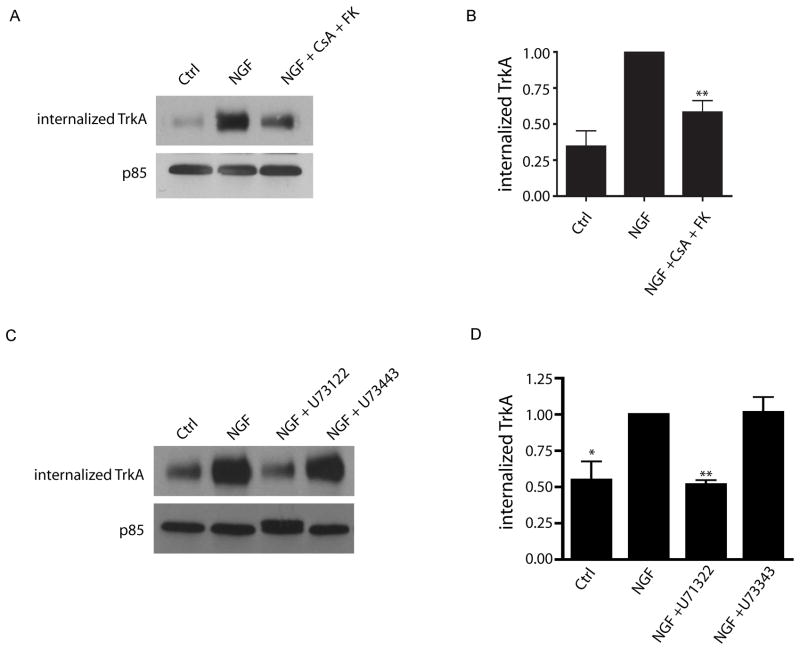
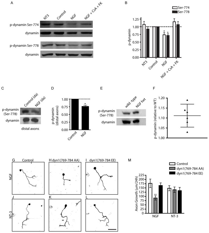
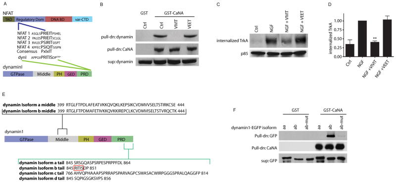
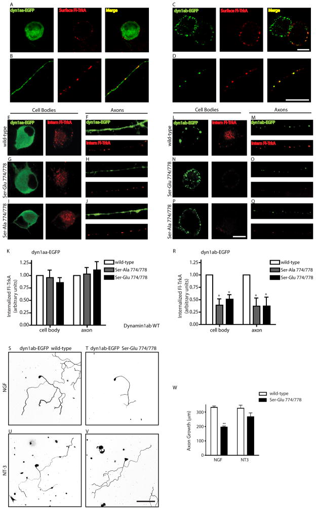
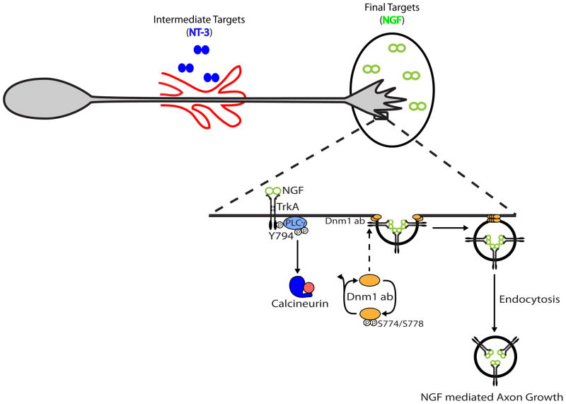
Endocytic events are critical for neuronal survival in response to target-derived neurotrophic cues, but whether local axon growth is mediated by endocytosis-dependent signaling mechanisms remains unclear. Here, we report that Nerve Growth Factor (NGF) promotes endocytosis of its TrkA receptors and axon growth by calcineurin-mediated dephosphorylation of the endocytic GTPase dynamin1. Conditional deletion of calcineurin in sympathetic neurons disrupts NGF-dependent innervation of peripheral target tissues. Calcineurin signaling is required locally in sympathetic axons to support NGF-mediated growth in a manner independent of transcription. We show that calcineurin associates with dynamin1 via a PxIxIT interaction motif found only in specific dynamin1 splice variants. PxIxIT-containing dynamin1 isoforms colocalize with surface TrkA receptors, and their phosphoregulation is selectively required for NGF-dependent TrkA internalization and axon growth in sympathetic neurons. Thus, NGF-dependent phosphoregulation of dynamin1 is a critical event coordinating neurotrophin receptor endocytosis and axonal growth.


Figures

Figure 1
Figure 1. Calcineurin is required for NGF, but not NT-3-mediated axon growth in sympathetic neurons
(A–L) Whole mount TH immunostaining shows reduced sympathetic fibers in target tissues in CaNB1fl/fl;Nestin-Cre mice as compared to CaNB1fl/fl controls, at E16.5 (heart: A–D), and E18.5 (heart: E–H and salivary glands: I–L). Higher magnification images are shown in the lower panels. Black arrows in I, J indicate sympathetic fibers approaching the salivary glands. Scale bar: 500 μm. (M–N) There are no differences in sympathetic chain organization between E16.5 CaNB1fl/fl (M) and CaNB1fl/fl;Nestin-Cre (N) mice. White arrow indicates TH-positive sympathetic fibers extending from sympathetic ganglia in both wild-type and mutant mice. Scale bar: 500 μm. (n=2 embryos for each genotype at E16.5, and at E18.5) (O) CaNB1fl/fl sympathetic neurons were infected with adenoviral vectors expressing Cre (Ad-Cre) or LacZ (Ad-LacZ). Neurotrophins were added only to distal axons (da). (P–S) Cre-mediated calcineurin deletion specifically decreases NGF, but not NT-3-mediated axon growth. Axons were stained with β-III-tubulin for visualization after quantification of axon growth. Scale bar, 80μm. (T) Quantification of axon growth in compartmentalized cultures over 0–8 hr and 0–24 hr, ** p<0.01, ***p<0.001. Results are mean ± SEM from n=5 experiments.
Figure 2
Figure 2. Calcineurin signaling is required in axons for NGF-mediated growth
(A–D) NGF-mediated axon growth is reduced by addition of calcineurin inhibitors (CsA+FK506) to distal axons (da) (C), but not cell bodies (cb) (D). NGF (100 ng/ml) was added only to distal axons. Axons were stained with β-III-tubulin for visualization. Scale bar, 320μm. (E) Quantification of NGF-mediated axon growth in compartmentalized cultures over 0–8 hr or 0–24 hr. * p<0.05, **p<0.01, n=4 experiments. (F–I) Calcineurin signaling is not required for NT-3-mediated axon growth. (J) Quantification of NT-3-mediated axon growth, n=4. (K) NGF but not NT-3 induces phosphorylation of TrkA on Tyr-794. Neuronal lysates were probed for phospho-TrkA (Y794). Immunoblots were reprobed for p85. (L) Densitometric quantification of phospho-TrkA (Y794). **p<0.01, n=3. (M) NGF treatment selectively promotes tyrosine phosphorylation of PLC-γ. Lysates were immunoprecipitated with anti-phospho-tyrosine and probed for PLC-γ. Supernatants were probed for p85. (N) Densitometric quantification of PLC-γ phosphorylation, ***p<0.001, n=4.
Figure 3
Figure 3. Calcineurin supports NGF-mediated axon growth in a transcription-independent manner
(A–C) NGF does not promote nuclear import of NFAT transcription factors in sympathetic neurons. NFAT immunostaining shows that NGF treatment (100 ng/ml, 30 min) does not induce nuclear localization of NFAT (B, arrow), while NFAT nuclear labeling is evident in neurons expressing CA-CaN (C, arrow). Neurons were also immunostained with β-III Tubulin and DAPI. Scale bar, 10μm. (D) NFAT-luciferase reporter assay shows that NGF and NT-3 do not activate NFAT-dependent transcription in sympathetic neurons. Neurons expressing CA-CaN show activation of NFAT-dependent transcription at 8 and 24 hr post-infection with NFAT-adenovirus, which is blocked by ActD (0.1 μg/ml). **p<0.01 and ***p<0.001. Results are means± SEM from 3 experiments. (E–I) Calcineurin signaling is required for NGF-mediated axon growth in the absence of transcription. NGF and calcineurin inhibitors (CsA+FK506) were added only to distal axons. ActD (0.1 μg/ml) was bath applied. Scale bar, 320μm. (I) Quantification of sympathetic axon growth in compartmentalized cultures over 0–8 hr and 0–24 hr after treatments described in (E–H). *p<0.05, **p<0.01, and ***p<0.001, n=3 experiments. (J) NFAT-luciferase assay shows that NGF does not activate NFAT-dependent transcription in DRG sensory neurons. DRG neurons expressing CA-CaN show robust activation of NFAT-dependent transcription. *p<0.05, **p<0.01, and ***p<0.001, n=5 experiments. (K–N) Calcineurin signaling is required for NGF-mediated axon growth in DRG neurons, in the absence of transcription. Scale bar, 320μm. (O) Quantification of DRG axon growth in compartmentalized cultures. *p<0.05, ** p<0.01, n=4.
Figure 4
Figure 4. PLC-γ and calcineurin mediate TrkA endocytosis
(A) Calcineurin activity is required for NGF-dependent internalization of TrkA receptors. Cell surface biotinylation and TrkA immunoblotting shows that NGF-dependent TrkA endocytosis is reduced by calcineurin inhibiton. (B) Densitometric quantification of internalized TrkA, **p<0.01, n=6. (C) TrkA endocytosis is dependent on PLC-γ activity. PLC-γ inhibitor (U73122) decreases NGF-dependent TrkA endocytosis. (D) Densitometric quantification of internalized TrkA, *p<0.05 and **p<0.01, n=4. In (A) and (C), supernatants were probed for p85 for normalization.
Figure 5
Figure 5. NGF promotes axon growth through dynamin dephosphorylation
(A) NGF stimulation results in dephosphorylation of dynamin1 in a calcineurin-dependent manner. Neuronal lysates were immunoblotted using phospho-Ser774 and phospho-Ser778 dynamin antibodies. Immunoblots were stripped and reprobed for total dynamin1. (B) Densitometric quantification of phospho-dynamin1 levels *p<0.05, n=6. (C) NGF stimulation results in dephosphorylation of dynamin1 (Ser 778) in distal axons. Immunoblots were reprobed for total dynamin1. (D) Densitometric quantification of phospho-dynamin1 (Ser778) in axons, *p<0.05, n=3. (E–F) NGF+/− mice have increased levels of phospho-dynamin1 in sympathetic axons in vivo. Salivary gland lysates from P0.5 wildtype and NGF+/− mice were immunoblotted using phospho-dynamin1 (Ser778) antibody. Immunoblots were reprobed for total dynamin1. (F) Densitometric quantification of phospho-dynamin1 (Ser778) after treatments as described in (E), represented as a scatter plot with 95% confidence intervals. n=6 pups for each genotype. (G) Dephosphorylation-dependent dynamin1 function is required for NGF-mediated axon growth. Introduction of dyn1(769-784 AA) (H) but not the dyn1(769-784 EE) (I) peptide decreased NGF-dependent axon growth over 24 hr. NT-3-mediated growth was unaffected by introduction of dyn1 phosphopeptides (J–L). Scale bar, 100μm. (M) Quantification of axon growth. **p<0.01, n=3.
Figure 6
Figure 6. Calcineurin-dynamin1 interaction is mediated by a PxIxIT motif found in specific dynamin1 isoforms
(A) Schematic of PxIxIT box consensus sequence found in the regulatory domain of NFAT (1–4) transcription factors and the PRITIS sequence in the proline-rich domain (PRD) of dynamin1. TAD is the transactivation domain, DNA BD is the DNA binding domain, var-CTD is the variable C-terminal domain for NFAT. PH is the pleckstrin homology domain and GED is the GTPase effector domain for dynamin. (B) Calcineurin-dynamin1 interaction is dependent on the PxIxIT motif. VIVIT peptide (a PxIxIT box mimic), but not a control VEET peptide, blocks association of CaNA with dynamin1. Pulldown with GST alone is shown as control. (C) Calcineurin-dynamin1 interaction via the PxIxIT motif is required for NGF-dependent TrkA internalization. Cell surface biotinylation assay shows that VIVIT, but not VEET treatment decreases NGF-dependent internalization of TrkA receptors. Supernatants were probed for p85. (D) Densitometric quantification of internalized TrkA, **p<0.01, n=4 (E, F) Calcineurin interaction is specific to dynamin1 variants with a PxIxIT box. (E) Schematic of dynamin1 splicing variants. Red box indicates xIxIS portion of the PRITIS box sequence, only present in b tail isoforms. (F) GST pull-down assays with HEK293 lysates show that calcineurin interacts with dynamin1ab via the PxIxIT box, but not dynamin1aa isoforms. HEK293 cells were transfected with dynamin1aa-EGFP, dynamin1ab-EGFP, or dynamin1ab-EGFP with PRITIS sequence mutated to ARATAA.
Figure 7
Figure 7. Phosphoregulation of dynamin1ab is required for TrkA endocytosis and axon growth
(A–D) Dynamin1aa isoform (A,B) shows diffuse cytoplasmic localization while dynamin1ab isoform (C,D) shows punctate localization in cell bodies and axons. FLAG immunostaining shows surface FLAG-TrkA receptors in cell bodies (A,C) and in axons (B,D). Scale bar, 10 μm. (E–J) Phosphoregulation of dynamin1aa is not required for NGF-dependent TrkA internalization. Neurons were transfected with FLAG-TrkA and either wild-type dynamin1aa-EGFP (E, F), Ser-Glu 774/778 dynamin1aa-EGFP (G,H), or Ser-Ala 774/778 dynamin1aa-EGFP (I,J). Cell bodies are shown in E, G, I. Axons are shown in F, H, J. Scale bar, 10μm. (K) Quantification of NGF-dependent TrkA internalization in cell bodies and axons. n=3. (L–Q) Phosphomutants of dynamin1ab disrupt NGF-dependent internalization of TrkA. Neurons were transfected with FLAG-TrkA and either wild-type dynamin1ab-EGFP (L,M), Ser-Glu 774/778 dynamin1ab-EGFP (N,O), or Ser-Ala 774/778 dynamin1ab-EGFP (P,Q). Cell bodies are shown in L, N and P. Axons are shown in M, O and Q. Scale bar, 10μm. (R) Quantification of internalized TrkA. *p<0.01, n=3. (S–V) Phosphoregulation of dynamin1ab is required for NGF-, but not NT-3-, dependent axon growth. NGF-mediated growth is blocked in sympathetic neurons expressing dynamin1ab-EGFP Ser-Glu 774/778 (T) as compared to wild-type dynamin1ab-EGFP (S). NT-3-mediated growth was unaffected (U,V). (W) Quantification of neurite length. **p<0.01, n=3. Scale bar: 50 μm.
Figure 8
Figure 8. NGF and NT-3 differentially employ calcineurin signaling to promote axonal growth in sympathetic neurons
NT-3 and NGF act upon a common TrkA receptor in sympathetic neurons to regulate distinct stages of axonal growth. NT-3 promotes proximal axon extension along the vasculature, whereas NGF is required for innervation of final peripheral targets. NGF-specific engagement of the PLC-γ effector pathway activates the calcium-responsive phosphatase, calcineurin. Calcineurin dephosphorylates PxIxIT motif-containing dynamin1 isoforms to promote TrkA endocytosis. Calcineurin-mediated endocytic trafficking of TrkA receptors signals locally in sympathetic nerve terminals to promote NGF-dependent axonal extension and branching within final target tissues.

References

    1. Anggono V, Smillie KJ, Graham ME, Valova VA, Cousin MA, Robinson PJ. Syndapin I is the phosphorylation-regulated dynamin I partner in synaptic vesicle endocytosis. Nat Neurosci. 2006;9:752–760. - PMC - PubMed
    1. Aramburu J, Garcia-Cozar F, Raghavan A, Okamura H, Rao A, Hogan PG. Selective inhibition of NFAT activation by a peptide spanning the calcineurin targeting site of NFAT. Mol Cell. 1998;1:627–637. - PubMed
    1. Aramburu J, Yaffe MB, Lopez-Rodriguez C, Cantley LC, Hogan PG, Rao A. Affinity-driven peptide selection of an NFAT inhibitor more selective than cyclosporin A. Science. 1999;285:2129–2133. - PubMed
    1. Ascano M, Richmond A, Borden P, Kuruvilla R. Axonal targeting of Trk receptors via transcytosis regulates sensitivity to neurotrophin responses. J Neurosci. 2009;29:11674–11685. - PMC - PubMed
    1. Beattie EC, Carroll RC, Yu X, Morishita W, Yasuda H, von Zastrow M, Malenka RC. Regulation of AMPA receptor endocytosis by a signaling mechanism shared with LTD. Nat Neurosci. 2000;3:1291–1300. - PubMed

Publication types