Comparative Study
. 2011 Jun 23;70(6):1218-25.
doi: 10.1016/j.neuron.2011.04.018.

Disrupted neural synchronization in toddlers with autism

Affiliations
Comparative Study

Disrupted neural synchronization in toddlers with autism

Ilan Dinstein et al. Neuron. .

Abstract

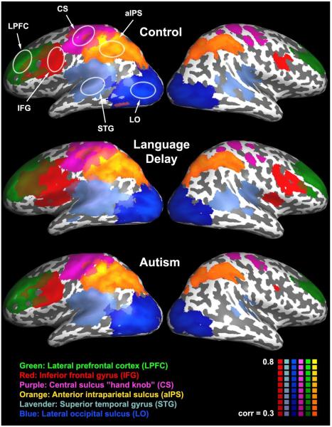
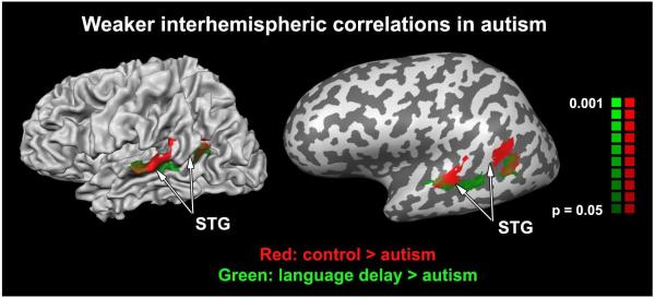
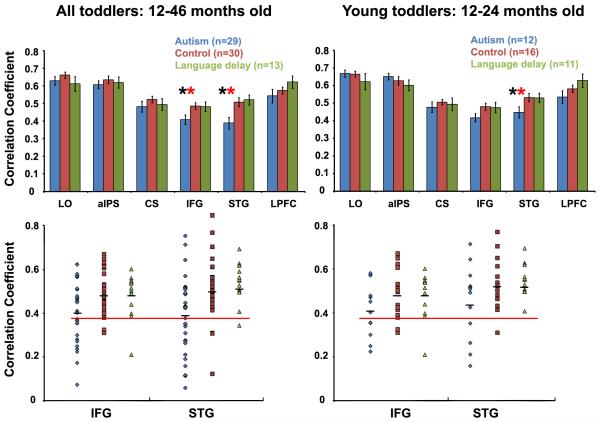
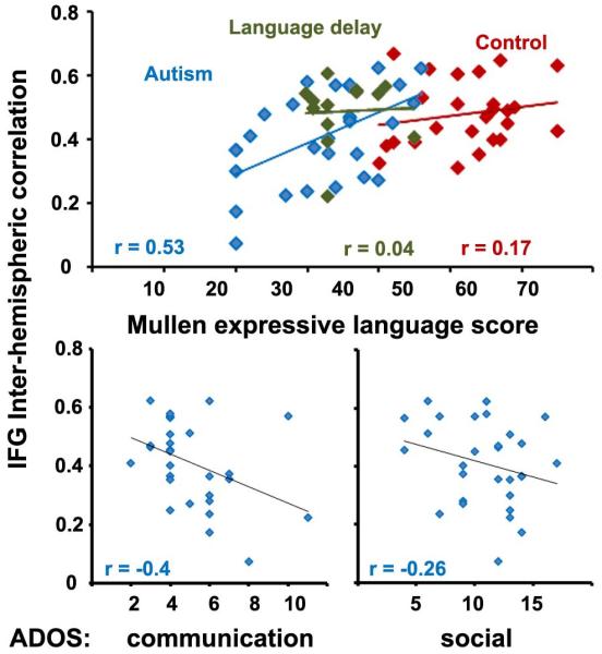
Autism is often described as a disorder of neural synchronization. However, it is unknown how early in development synchronization abnormalities emerge and whether they are related to the development of early autistic behavioral symptoms. Here, we show that disrupted synchronization is evident in the spontaneous cortical activity of naturally sleeping toddlers with autism, but not in toddlers with language delay or typical development. Toddlers with autism exhibited significantly weaker interhemispheric synchronization (i.e., weak "functional connectivity" across the two hemispheres) in putative language areas. The strength of synchronization was positively correlated with verbal ability and negatively correlated with autism severity, and it enabled identification of the majority of autistic toddlers (72%) with high accuracy (84%). Disrupted cortical synchronization, therefore, appears to be a notable characteristic of autism neurophysiology that is evident at very early stages of autism development.


Figures

Figure 1
Figure 1
Correlation maps averaged across toddlers from the typically-developing (top), language delay (middle), and autism (bottom) groups. fMRI activity during natural sleep was sampled in six left hemisphere “seed” locations outlined by white ellipses: Lateral prefrontal cortex (LPLC), inferior frontal gyrus (IFG), “hand knob” area of central sulcus (CS), anterior intraparietal sulcus (aIPS), superior temporal gyrus (STG), and lateral occipital sulcus (LO). Each color represents voxels that exhibited strong correlation (above 0.3) with a particular seed. Note the spatial selectivity of the correlations in all groups. Only voxels located close to the seed’s location in the left hemisphere and the corresponding contralateral location in the right hemisphere exhibited strong correlation values.
Figure 2
Figure 2
Voxels exhibiting weaker inter-hemispheric correlations in the autism group as compared with the typically developing (red) and language delay (green) groups. The two independent comparison maps are overlaid on a folded (left) and inflated (right) left hemisphere of a single individual. Significantly weaker inter-hemispheric correlation was apparent in STG voxels in both comparisons. No voxels exhibited stronger inter-hemispheric correlation in children with autism. STG – superior temporal gyrus.
Figure 3
Figure 3
Inter-hemispheric correlation strength between right and left ROIs in the autism (blue), typically developing (red), and language delay (green) groups when considering all subjects (left panels) or only the younger toddlers (right panels). Top panels: Average correlation strength in each toddler group for each of the six examined ROIs. The autism group exhibited significantly weaker inter-hemispheric correlation (p < 0.05) only in putative language areas (IFG and STG). When comparing younger toddler groups IFG correlation difference was almost significant (p < 0.07). Error bars: standard error across subjects. Black asterisk: significant difference between autism and control groups. Red asterisk: significant difference between autism and language delay groups. Bottom panels: Single subject correlation values in IFG and STG. The majority of toddlers with autism, but only a small minority of control (red) and language delay (green) toddlers, exhibited IFG or STG correlation values below 0.38 (red line). Black lines: mean correlation across the group.
Figure 4
Figure 4
Inter-hemispheric correlation in IFG and verbal ability (top) or autism severity (bottom). Toddlers with autism (blue) showed a significant positive correlation between inter-hemispheric correlation value and their expressive language ability as measured by the Mullen test (top), while typically developing (red) and language delayed toddlers (green) did not. Toddlers with autism exhibited a significant negative correlation between inter-hemispheric correlation and the ADOS communications score (left) while exhibiting a negative trend with the ADOS social scores (right).

References

    1. Alexander AL, Lee JE, Lazar M, Boudos R, DuBray MB, Oakes TR, Miller JN, Lu J, Jeong EK, McMahon WM, et al. Diffusion tensor imaging of the corpus callosum in Autism. Neuroimage. 2007;34:61–73. - PubMed
    1. Anderson JS, Druzgal TJ, Froehlich A, Dubray MB, Lange N, Alexander AL, Abildskov T, Nielsen JA, Cariello AN, Cooperrider JR, et al. Decreased Interhemispheric Functional Connectivity in Autism. Cereb Cortex. 2010 - PMC - PubMed
    1. Belmonte MK, Allen G, Beckel-Mitchener A, Boulanger LM, Carper RA, Webb SJ. Autism and abnormal development of brain connectivity. J Neurosci. 2004;24:9228–9231. - PMC - PubMed
    1. Ben Bashat D, Kronfeld-Duenias V, Zachor DA, Ekstein PM, Hendler T, Tarrasch R, Even A, Levy Y, Ben Sira L. Accelerated maturation of white matter in young children with autism: a high b value DWI study. Neuroimage. 2007;37:40–47. - PubMed
    1. Birn RM, Diamond JB, Smith MA, Bandettini PA. Separating respiratory-variation-related fluctuations from neuronal-activity-related fluctuations in fMRI. Neuroimage. 2006;31:1536–1548. - PubMed

Publication types

MeSH terms

LinkOut - more resources