. 2011 Aug;179(2):915-30.
doi: 10.1016/j.ajpath.2011.04.008. Epub 2011 May 31.

Follistatin improves skeletal muscle healing after injury and disease through an interaction with muscle regeneration, angiogenesis, and fibrosis

Affiliations

Follistatin improves skeletal muscle healing after injury and disease through an interaction with muscle regeneration, angiogenesis, and fibrosis

Jinhong Zhu et al. Am J Pathol. 2011 Aug.

Abstract

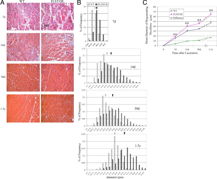
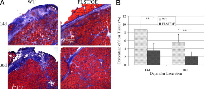
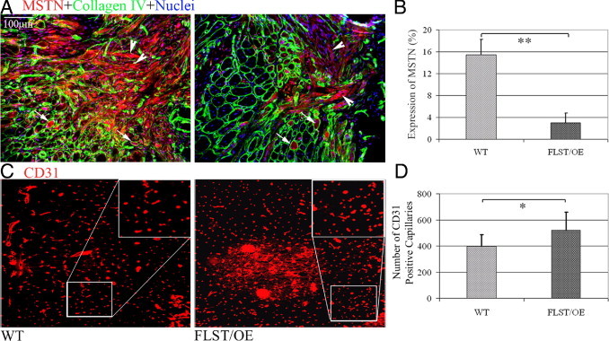
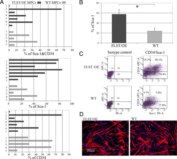
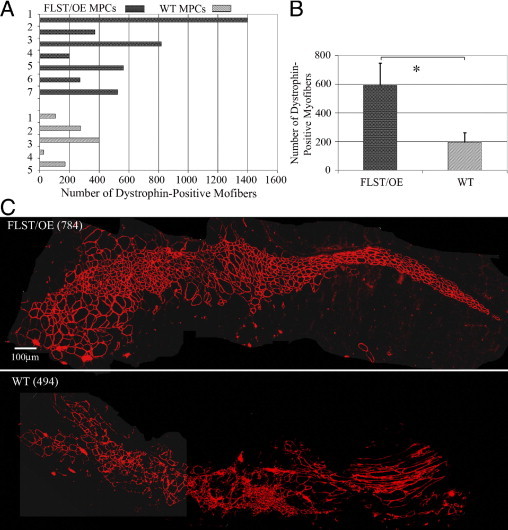
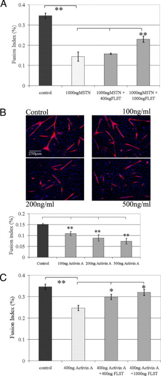
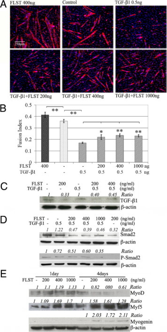
Recovery from skeletal muscle injury is often incomplete because of the formation of fibrosis and inadequate myofiber regeneration; therefore, injured muscle could benefit significantly from therapies that both stimulate muscle regeneration and inhibit fibrosis. To this end, we focused on blocking myostatin, a member of the transforming growth factor-β superfamily and a negative regulator of muscle regeneration, with the myostatin antagonist follistatin. In vivo, follistatin-overexpressing transgenic mice underwent significantly greater myofiber regeneration and had less fibrosis formation compared with wild-type mice after skeletal muscle injury. Follistatin's mode of action is likely due to its ability to block myostatin and enhance neovacularization. Furthermore, muscle progenitor cells isolated from follistatin-overexpressing mice were significantly superior to muscle progenitors isolated from wild-type mice at regenerating dystrophin-positive myofibers when transplanted into the skeletal muscle of dystrophic mdx/severe combined immunodeficiency mice. In vitro, follistatin stimulated myoblasts to express MyoD, Myf5, and myogenin, which are myogenic transcription factors that promote myogenic differentiation. Moreover, follistatin's ability to enhance muscle differentiation is at least partially due to its ability to block myostatin, activin A, and transforming growth factor-β1, all of which are negative regulators of muscle cell differentiation. The findings of this study suggest that follistatin is a promising agent for improving skeletal muscle healing after injury and muscle diseases, such as the muscular dystrophies.


Figures

Figure 1
Figure 1
Injured follistatin-overexpressing (FLST/OE) skeletal muscles showed accelerated regeneration compared with their WT counterparts. A: H&E staining of cross-sections of injured WT and FLST/OE skeletal muscle at 7, 14, and 30 days and 1.5 years after laceration injury. The myofibers and nuclei stained red and black, respectively. Original magnification, ×200. Regenerating myofibers are characterized by centralized nuclei. Black scale bar represents 100 μm. B: Distribution of diameters of regenerating myofibers in WT and FLST/OE skeletal muscle 7 (n = 3), 14 (n = 8), and 30 (n = 8) days and 1.5 years (n = 4) after injury. Gray bars represent myofibers from WT mice, whereas black bars represent myofibers from FLST/OE mice. Moreover, the gray arrowheads indicate mean diameters of regenerating fibers in WT muscle, whereas black arrowheads indicate mean diameters of regenerating fibers in FLST/OE muscle. C: Regenerating myofiber diameter quantifications. The smallest diameters of more than 300 nonadjacent myofibers per muscle were measured using Northern Eclipse software. The mean diameters of regenerating fibers were shown to increase in both WT and FLST/OE muscles over time after injury; however, the mean diameters of the FLST/OE fibers were significantly greater at all time points (*P < 0.05, **P < 0.01).
Figure 2
Figure 2
Fibrosis formation in the injured follistatin-overexpressing (FLST/OE) muscle was reduced when compared with the injured WT muscle. Masson's trichrome staining was performed on sections of injured FLST/OE and WT muscle (myofibers in red; fibrosis in blue). A: Representative images of injured FLST/OE and WT muscle at 14 (n = 8) and 30 (n = 8) days after injury. Original magnification, ×100. There was significantly less fibrosis observed in the injured FLST/OE muscles than the WT muscles. B: Injured FLST/OE muscles developed significantly less fibrosis than did injured WT muscles (**P < 0.01).
Figure 3
Figure 3
Decreased myostatin (MSTN) expression and increased angiogenesis in injured follistatin-overexpressing (FLST/OE) skeletal muscles. A: IHC analysis was performed to detect MSTN (red) and collagen type IV (green) expression in injured WT and FLST/OE muscle. Collagen type IV was used to highlight the basal lamina of myofibers, including necrotic, intact, and regenerating myofibers. MSTN-positive signals were seen within some of the regenerating myofibers surrounded by the basal lamina (arrows) and some expression outside the basal lamina (arrowheads). Original magnification, ×200. Injured FLST/OE muscles contained less MSTN staining than did injured WT muscles. B: When we measured the relative MSTN positive signals and areas, we found that there was significantly more MSTN expression detected in the injured WT muscles than in injured FLST/OE muscles. C: CD31, an endothelial cell marker, was used to stain capillary-like structures in the injured muscles. Original magnification, ×200. D: There were a significantly greater number of CD31+ signals present in the injured FLST/OE muscles than in the injured WT muscles (n = 8) (*P < 0.05, **P < 0.01).
Figure 4
Figure 4
Characterization of MPCs. Seven follistatin-overexpressing (FLST/OE) and five WT MPC populations were examined for Sca-1 expression, CD34 expression, and in vitro myogenic differentiation. A: Histograms showing wild variability in the percentages of Sca-1+ and CD34+, Sca-1+, and CD34+ expressing cells among the MPC populations. B: Quantitation revealed a significant increase in the Sca-1+ fraction in the FLST/OE MPC populations compared with the WT MPC populations. C: Images on the left are isotype controls; images on the right are representative images of a flow cytometry dot plot showing that FLST/OE MPC populations consist of a larger proportion of Sca-1+ cells than the WT MPC populations (46.5% versus 24%). APC-A indicates allophycocyanin-area; PE-A, phycoerythrin area. D: Both FLST/OE and WT MPC populations underwent myogenic differentiation as labeled by MyHC (red) and DAPI (blue). Original magnification, ×100.
Figure 5
Figure 5
Follistatin-overexpressing MPCs (FLST/OE MPCs) regenerated skeletal muscle more efficiently than WT MPCs, when transplanted into the GMs of mdx/SCID mice. A: Quantitation of engraftment in terms of the number of dystrophin-positive fibers regenerated by the FLST/OE and WT MPC populations. B: The overall mean ± SEM number of dystrophin-positive myofibers was significantly greater for the FLST/OE MPCs (592.8 ± 154.9; n = 7 FLST/OE MPC populations; four muscles per population) than for the WT MPC populations (195.6 ± 65.4; n = 5 WT MPC populations; four muscles per population; **P = 0.023, Student's t-test). C: Representative engraftments showed that the transplanted MPCs regenerated dystrophin-positive myofibers (red) within dystrophic muscle. FLST/OE MPCs produced more dystrophin-positive myofibers than did WT MPCs (*P < 0.05, **P < 0.01). Original magnification, ×200.
Figure 6
Figure 6
Interactions between follistatin (FLST) and myostatin (MSTN) and FLST and activin A. A: As indicated by the fusion index (the ratio of nuclei in myotubes to total nuclei), MSTN significantly inhibited C2C12 myoblast differentiation, but FLST counteracted MSTN and attenuated its inhibition of cellular differentiation. B: Without intervention, C2C12 myoblasts underwent myogenic differentiation in low serum medium as evidenced with MyHC (red) and DAPI (blue). Activin A significantly reduced muscle cell differentiation and the formation of myotubes in cell culture. Original magnification, ×100. C: FLST could neutralize the inhibitory effect of activin A on myoblast differentiation as shown by FLST induced-restoration of differentiation (n = 3; *P < 0.05, **P < 0.01).
Figure 7
Figure 7
Follistatin (FLST) neutralized TGF-β1's activity on C2C12 myoblasts. A: Exemplary pictures of differentiation of C2C12 myoblasts treated by FLST alone, TGF-β1, and combinations of FLST and TGF-β1. Original magnification, ×100. Myotubes were visualized with MyHC (red) and DAPI (nuclei, blue). B: Our quantitative results showed that TGF-β1 significantly inhibited myogenic differentiation of C2C12 myoblasts. FLST was able to reverse TGF-β1–inhibited myogenic differentiation (n = 3). C: Western blot results showed that FLST decreased TGF-β1 expression in C2C12 myoblasts with or without the presence of exogenous TGF-β1. D: FLST also reduced the phosphorylation of SMAD2. E: FLST stimulated the expressions of the myogenic regulatory factors, MyoD, Myf5, and myogenin by myoblasts; the quantification of the Western blots was indicated as normalized ratio of proteins of interest to β-actin, whereas controls were referred to as 1 (*P < 0.05, **P < 0.01).
Figure 8
Figure 8
AAV2-MPRO improves skeletal muscle healing at 4 weeks after injury. A: Masson's trichrome staining shows that fibrotic tissue (blue) exists at the muscle injury site. B: Fibrosis in the AAV2-MPRO–treated injured muscles was significantly less than the untreated control. Original magnification, ×200. C: Collagen IV (green) and MPRO (red) double-immunostaining shows a strong MPRO signal in the cytoplasm of the fibers of AAV2-MPRO transduced muscle. D: AAV2-MPRO transduced GMs gained significantly more weight than the GM controls. E: H&E staining revealed muscle hypertrophy in the AAV2-MPRO–treated injured muscles. F: A significant increase in the average diameter of the regenerating myofibers was observed in the AAV2-MPRO injected muscles when compared with the controls (**P < 0.01). Original magnification, ×100. G: Frequency histograms show the distribution of regenerating myofibers in the AAV-MPRO–treated and control muscles. Scale bar = 100 μm.

Similar articles

Cited by

References

    1. Border W.A., Noble N.A. Transforming growth factor beta in tissue fibrosis. N Engl J Med. 1994;331:1286–1292. - PubMed
    1. Lijnen P.J., Petrov V.V., Fagard R.H. Induction of cardiac fibrosis by transforming growth factor-beta(1) Mol Genet Metab. 2000;71:418–435. - PubMed
    1. Waltenberger J., Lundin L., Oberg K., Wilander E., Miyazono K., Heldin C.H., Funa K. Involvement of transforming growth factor-beta in the formation of fibrotic lesions in carcinoid heart disease. Am J Pathol. 1993;142:71–78. - PMC - PubMed
    1. Yamamoto T., Noble N.A., Miller D.E., Border W.A. Sustained expression of TGF-beta 1 underlies development of progressive kidney fibrosis. Kidney Int. 1994;45:916–927. - PubMed
    1. Li Y., Foster W., Deasy B.M., Chan Y., Prisk V., Tang Y., Cummins J., Huard J. Transforming growth factor-beta1 induces the differentiation of myogenic cells into fibrotic cells in injured skeletal muscle: a key event in muscle fibrogenesis. Am J Pathol. 2004;164:1007–1019. - PMC - PubMed

Publication types