. 2011 Aug;141(2):448-59.
doi: 10.1053/j.gastro.2011.06.003. Epub 2011 Jun 12.

Notch: architect, landscaper, and guardian of the intestine

Affiliations

Notch: architect, landscaper, and guardian of the intestine

Marc Vooijs et al. Gastroenterology. 2011 Aug.

Abstract

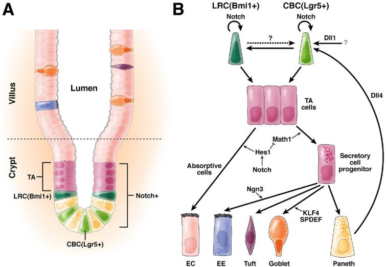
In the past decade, enormous progress has been made in understanding the role of stem cells in physiologic tissue renewal and in pathologic processes such as cancer. These findings have shed light on the identity and biological properties of such cells and the intrinsic and extrinsic signals that balance stem cell self-renewal with differentiation. With its astonishing self-renewal capacity, the intestinal epithelium has provided a unique model to study stem cell biology, lineage specification, and cancer. Here we review the role of Notch signaling in physiologic cell renewal and differentiation in the intestine as well as during its malignant transformation.


Figures

Figure 1
Figure 1. The mammalian small intestine and role of Notch in ISC renewal.
(A) Two populations of stem cells, CBCs and label retaining cells (LRCs), characterized by Lgr5 and Bmi1 expression, respectively, are located at the bottom of the crypt. Their daughters, the TA cells, undergo rapid proliferation as they move upwards. Terminal cell differentiation starts at about the upper third of the crypt and gives rise to 5 major cell types: the absorptive ECs and the secretory cell types composed of EE cells, tuft cells, goblet cells, and Paneth cells. Whereas most differentiated cells move and home into the villi, Paneth cells move back into the bottom of the crypt after their birth to form the niche to orchestrate ISC renewal and differentiation. (B) The relationship between the LRC and CBC stem cell populations is not very clear. It is possible that they could give rise to each other (dashed arrows). Although both populations produce TA cells, it has been proposed that CBCs may represent a rapidly cycling population (thick arrow) whereas LRC is a slow cycling one (thin arrow). Notch may be required for the maintenance of both. Although Paneth cells express Dll4 ligand, it remains unclear which cells provide the Dll1 ligands, which play a dominant role in sustaining stem cell self-renewal as well. During cell fate specification, Notch promotes the EC fate and inhibits the secretory cell fates through down-regulation of Math1, a master transcription factor for all secretory cell fates. Math1 controls secretory commitment by activation of cell type-specific transcription factors. For example, Ngn3 specifies the EE cell fates while KLF4 and SPDEF goblet cells.
Figure 2
Figure 2. The core components of the Notch signaling pathway.
(A) Typical domain structure of the vertebrate Notch receptor and ligands. Jagged and Delta ligands share from N-terminus to C-terminus the Delta/Serrate/LAG-2 domain (DSL), the Delta and OSM-11-like protein domain (DOS), a variable number of EGF repeats, transmembrane domain (TM), nuclear localization signal (NLS), and PSD95/Dlga/Zo-1domain (PDZ). Jagged1 and 2 also have a cysteine-rich domain (CR). Notch receptors have 24 to 36 EGF repeats, which mediate ligand interaction (11 and 12, shaded in pink, are in direct contact with the DOS domain of the ligands). Following these EGF repeats are 3 cysteine-rich Lin-12/Notch repeats (LNRs), a heterodimerization domain (HD), transmembrane domain (TD), the RBP-Jκ association module (RAM), 7 NLS-flanked ankyrin repeats (ANK), and the proline/glutamic acid/serine/threonine-rich degradation motif (PEST). (B) Key steps involved in the activation of the Notch signaling pathway. In the absence of Notch activity, RBP-Jκ binds to the promoters of Notch target genes such as Hes and Hey and recruits corepressors to repress transcription. Newly synthesized Notch receptor experiences a series of posttranslational modifications in the ER and Golgi body before reaching the cell surface, including the glycosylation by O-fucose-transferase (Ofut1) and Fringe as well as the S1 cleavage by furin-like convertases. In contrast, the ligands are ubiquitinated in the signaling-sending cell by ubiquitin ligases such as Mib-1, endocytosed, and then recycled back to the cell membrane. Trans-interactions between the Notch receptor and its ligands is mediated by EGF repeat 11-12 of the receptor and the DOS domain of the ligands followed by transendocytosis of the ligand. This causes unfolding of the LNR/HD domain exposing the S2 cleavage site a substrate for the ADAM (A Disintegrin And Metalloprotease) metalloproteases. S2 cleavage leads to shedding of the extracellular domain of the receptor and turns it into the substrate for the γ-secretase complex, which catalyzes the cleavage at the S3 site within the Notch transmembrane domain. This leads to the release of the NICD from the cell membrane and its subsequent translocation into the nucleus, where it displaces corepressor bound to RBP-Jκ and recruits coactivators such as Mastermind and p300 to activate gene expression.
Figure 3
Figure 3. Drosophila posterior midgut and role of Notch in ISC renewal.
(A) The Drosophila posterior midgut is a relatively simple pseudostratified epithelium, with only 2 differentiated cell types: ECs and EE cells. In addition, ISCs and their nonproliferative daughters, EBs, are scattered in between. (B) Two populations of ISCs exist: one with high Delta expression (ISC [DlHi]) and one with no or low Delta expression (ISC [DlLo]). When they divide, both give one daughter ISC and one daughter EB cell. However, the daughter ISC from ISC (DlHi) will activate a high level of Notch signaling in its sister EB cells, which drives its differentiation into EC cells; in contrast, the daughter ISC from ISC (DlLo) elicits a low level of Notch signaling in its sister EB cells, which will differentiate into EE cells. Thus, different levels of Notch activation elicit different cell fates.

References

    1. Vries RG, Huch M, Clevers H. Stem cells and cancer of the stomach and intestine. Mol Oncol. 2010;4:373–384. - PMC - PubMed
    1. Sancho E, Batlle E, Clevers H. Signaling pathways in intestinal development and cancer. Annu Rev Cell Dev Biol. 2004;20:695–723. - PubMed
    1. Cheng H, Bjerknes M. Cell production in mouse intestinal epithelium measured by stathmokinetic flow cytometry and Coulter particle counting. Anat Rec. 1983;207:427–434. - PubMed
    1. Totafurno J, Bjerknes M, Cheng H. The crypt cycle. Crypt and villus production in the adult intestinal epithelium. Biophys J. 1987;52:279–94. - PMC - PubMed
    1. Gerbe F, van Es JH, Makrini L, et al. Distinct ATOH1 and Neurog3 requirements define tuft cells as a new secretory cell type in the intestinal epithelium. J Cell Biol. 2011;192:767–780. - PMC - PubMed

Publication types