

Intestinal lymphatic transport for drug delivery
- PMID: 21689702
- PMCID: PMC7126116
- DOI: 10.1016/j.addr.2011.05.019
Intestinal lymphatic transport for drug delivery
Abstract
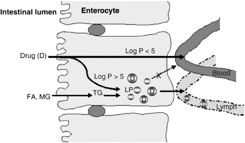
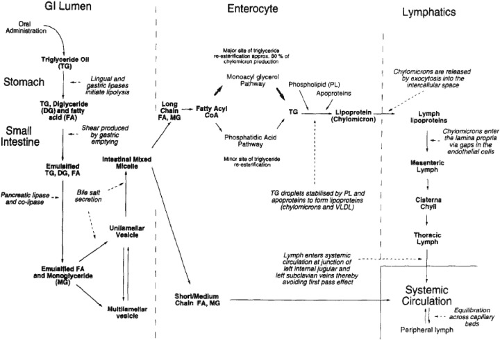
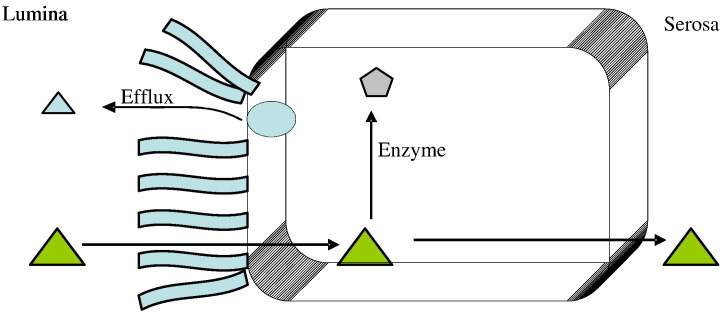
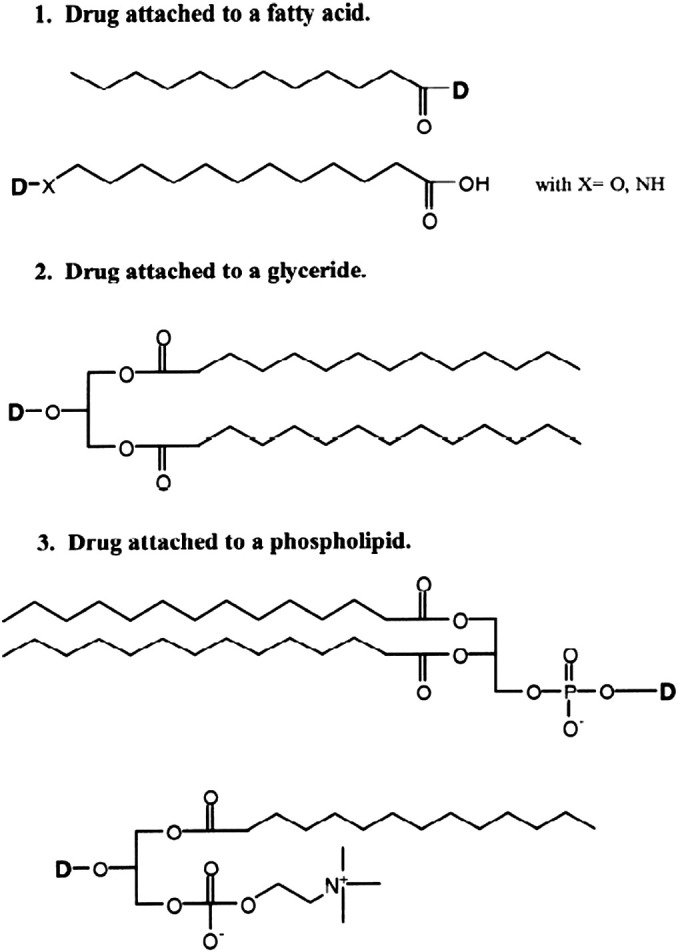
Intestinal lymphatic transport has been shown to be an absorptive pathway following oral administration of lipids and an increasing number of lipophilic drugs, which once absorbed, diffuse across the intestinal enterocyte and while in transit associate with secretable enterocyte lipoproteins. The chylomicron-associated drug is then secreted from the enterocyte into the lymphatic circulation, rather than the portal circulation, thus avoiding the metabolically-active liver, but still ultimately returning to the systemic circulation. Because of this parallel and potentially alternative absorptive pathway, first-pass metabolism can be reduced while increasing lymphatic drug exposure, which opens the potential for novel therapeutic modalities and allows the implementation of lipid-based drug delivery systems. This review discusses the physiological features of the lymphatics, enterocyte uptake and metabolism, links between drug transport and lipid digestion/re-acylation, experimental model (in vivo, in vitro, and in silico) of lymphatic transport, and the design of lipid- or prodrug-based drug delivery systems for enhancing lymphatic drug transport.
Copyright © 2011 Elsevier B.V. All rights reserved.
Figures
References
-
- Charman W.N., Stella V.J. Estimating the maximum potential for intestinal lymphatic transport of lipophillic drug molecules. Int. J. Pharm. 1986;34:175–178.
-
- Nordskog B.K., Phan C.T., Nutting D.F., Tso P. An examination of the factors affecting intestinal lymphatic transport of dietary lipids. Adv. Drug Deliv. Rev. 2001;50:21–44. - PubMed
-
- Cense H.A., van Eijck C.H., Tilanus H.W. New insights in the lymphatic spread of oesophageal cancer and its implications for the extent of surgical resection. Best Pract. Res. Clin. Gastroenterol. 2006;20:893–906. - PubMed
-
- Arya M., Bott S.R., Shergill I.S., Ahmed H.U., Williamson M., Patel H.R. The metastatic cascade in prostate cancer. Surg. Oncol. 2006;15:117–128. - PubMed
Publication types
MeSH terms
Substances
LinkOut - more resources
Full Text Sources
Other Literature Sources
