. 2011 Nov 1;92(2):199-208.
doi: 10.1093/cvr/cvr181. Epub 2011 Jun 20.

Enhanced K(+)-channel-mediated endothelium-dependent local and conducted dilation of small mesenteric arteries from ApoE(-/-) mice

Affiliations

Enhanced K(+)-channel-mediated endothelium-dependent local and conducted dilation of small mesenteric arteries from ApoE(-/-) mice

Timea Beleznai et al. Cardiovasc Res. .

Abstract

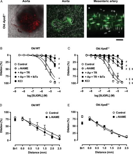
Aims: Agonists that evoke smooth muscle cell hyperpolarization have the potential to stimulate both local and conducted dilation. We investigated whether the endothelium-dependent vasodilators acetylcholine (ACh) and SLIGRL stimulated conducted dilation and whether this was altered by deficiency in apolipoprotein E (ApoE(-/-)).

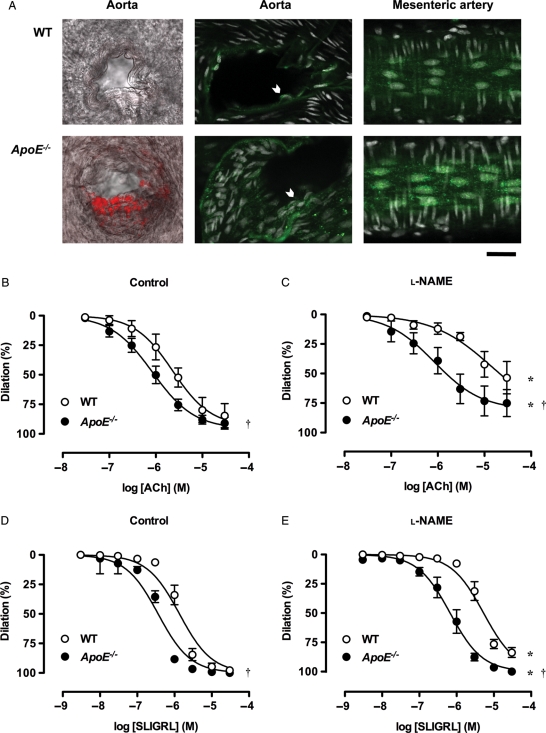
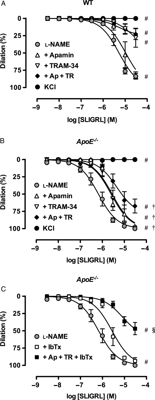
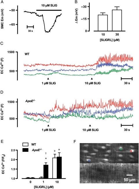
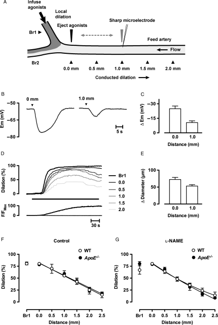
Methods and results: Isolated mesenteric arteries were cannulated, pressurized, and precontracted with phenylephrine. Agonists were either added to the bath to study local dilation or were restricted to one end of arteries to study conducted dilation. An enhanced sensitivity to both ACh and SLIGRL was observed in mesenteric arteries from ApoE(-/-) mice compared with wild-type controls. Inhibition of nitric oxide (NO) synthase blocked ACh responses, but had no effect on maximum dilation to SLIGRL. SLIGRL increased endothelial cell Ca(2+), hyperpolarized smooth muscle cells, and fully dilated arteries. The NO-independent dilation to SLIGRL was blocked with high [KCl] or Ca(2+)-activated K(+)-channel blockers. The hyperpolarization and dilation to SLIGRL passed through the artery to at least 2.5 mm upstream. The conducted dilation was not affected by a deficit in ApoE and could also be stimulated by ACh, suggesting NO itself could stimulate conducted dilation.

Conclusion: In small mesenteric arteries of ApoE(-/-) mice, NO-independent dilation is enhanced. Since both NO-dependent and -independent pathways can stimulate local and conducted dilation, the potential for reducing vascular resistance is improved in these vessels.


Figures

Figure 1
Figure 1
EDH-type dilation is augmented in mesenteric arteries from young ApoE−/− mice. (A) Visualization of atherosclerotic plaques (left and middle panels). Confocal micrographs of oil red O (red) and Mac-3 (green) staining in aortic branch points from WT and ApoE−/− mice. Plaque formation was only evident in the latter, although punctate intracellular staining for Mac-3 was evident in all endothelial cells (chevrons) (right panels). Images of endothelial cells in isolated, triple-cannulated and pressurized mesenteric arteries from WT and ApoE−/− mice. Mac-3 staining increased in the perinuclear region from ApoE−/− mice, but no plaques were observed using any method, either in the feed artery, or at the bifurcation. Nuclei were stained with propidium iodide (white). Bar = 30 µm. Images are representative of at least three arteries. (BE) Dilation to endothelium-dependent agonists. Cumulative concentration response curves to ACh and SLIGRL under control conditions (B and D) and in the presence of 100 µM l-NAME (C and E) in mesenteric arteries from WT and ApoE−/− mice (n = 3–14). *P< 0.05 compared with the control response of the same strain and age; P< 0.05 compared with WT.
Figure 2
Figure 2
K+-channels activated during EDH-type dilation in mesenteric arteries. Cumulative concentration response curves to SLIGRL in mouse mesenteric arteries from young WT (A) and ApoE−/− (B and C) mice. l-NAME (100 µM) alone was compared with the additional presence of apamin (1 µM), TRAM-34 (1 µM) or IbTx (0.1 µM), and various combinations (n = 3–14). These responses were compared with arteries precontracted with 45 mM KCl (n = 3). #P< 0.05 compared with l-NAME; P< 0.05 compared with WT; §P< 0.05 compared with l-N + Ap + TR in ApoE−/−.
Figure 3
Figure 3
Changes in membrane potential and endothelial cell Ca2+ in response to SLIGRL in pressurized mesenteric arteries from young mice. (A) Time course of hyperpolarization in response to 10 µM SLIGRL in mesenteric arteries. SLIGRL was added to the superfusion solution reservoir, indicated by the bar. Note there was a short delay before SLIGRL reached the artery. (B) Summary of the peak increase in membrane potential evoked by 10 µM (n = 7) and 30 µM (n = 3) SLIGRL in arteries from WT mice. (C–D) Time course of endothelial cell Ca2+ in response to 1 and 10 µM SLIGRL in an artery from a WT (C) and ApoE−/− (D) mouse. Each colour represents the average fluorescence intensity (F) in endothelial cells. The responses were asynchronous between cells. (E) Summary of changes in endothelial cell [Ca2+]i following addition of 1 and 10 µM SLIGRL (F) relative to basal intensity (F0) in arteries from WT and ApoE−/− mice (n = 4–5). *P< 0.05 compared with the control response (0 µM SLIGRL) of the same strain; P< 0.05 compared with WT. (F) Micrographs of the artery used to generate the data for D, showing the regions used for analysis (colours correspond to traces in D). A z-series was obtained from −10 µm (into smooth muscle cells) to +5 µm (into lumen) at 0.5 µm intervals from the plane of Ca2+-signal acquisition. The artery was loaded with Oregon Green® 488 BAPTA-1 (top panel) and exposed to 10 µM Alexa Fluor® 633 hydrazide (bottom panel) to show the internal elastic lamina (IEL) and other structures. The entire z-series was averaged to generate each image, and shows clear staining of endothelial cells and only very weak staining of smooth muscle cells. In general, the IEL could be used to monitor changes in focus during Ca2+-signal acquisition.
Figure 4
Figure 4
SLIGRL evokes conducted hyperpolarization and dilation in pressurized mesenteric arteries from young mice. (A) Schematic diagram of artery for studying conducted dilation. For electrophysiology, a sharp microelectrode was used to impale a smooth muscle cell to record membrane potential, and the agonist delivered by pressure ejection either locally or 1.0 mm downstream from the recording electrode. For luminal perfusion of agonists to study conducted dilation, arteries were triple cannulated to allow a syringe pump to deliver SLIGRL with a fluorescent indicator (dark grey) through the lumen of Branch 1 (Br1). Continuous flow through the Feed artery prevented upstream diffusion of agonist, but did not in itself stimulate dilation. The entire field of view was visible to simultaneously measure diameter in Branch 1 (Br1) and the positions along the Feed artery (0.0–2.5 mm upstream). (B) Time course of local (0.0 mm) and conducted (1.0 mm) hyperpolarization to a 100 ms pulse of SLIGRL delivered to the outside of a pressurized artery with a micropipette. Summary data from mesenteric arteries cannulated from three young WT mice are shown in C. (D) Time course of local and conducted dilation to SLIGRL. SLIGRL (30 µM) together with carboxyfluorescein were infused into Branch 1 (Br1) for the period indicated by the bar. Dilation and changes in fluorescence in Br1 and at 0.5 mm intervals along the vessel (0.0–2.5 mm) were measured simultaneously. (E) Summary of the changes in diameter at the 0.0 and 1.0 mm sites under control conditions in WT animals. Maximum diameter was 253 ± 16 µm (n = 7). Summary of local (Br1) and conducted dilation (0.0–2.5 mm) in each strain of mouse under control conditions (F, n = 3–8), and in arteries incubated with 100 µM l-NAME (G, n = 3–8).
Figure 5
Figure 5
Comparison of conducted dilation in pressurized mesenteric arteries from young mice. (A) Responses to ACh (10 µM, n = 4) in the absence (Control) and presence of 100 µM l-NAME. See Figure 4 for details. (B) The time courses of all conducted dilation experiments were analysed at points where the dilation at 0.0 mm was near 20, 40, 60, and 80% of maximum diameter, and the corresponding simultaneous dilation at the 1.0 site was plotted.
Figure 6
Figure 6
Arterial structure and function in older mice. (A, left and middle panel). Confocal micrographs of oil red O (red) and Mac-3 (green) staining in aortic branch points from old ApoE−/− mice (right panel). Images of endothelial cells in a mesenteric artery from and old ApoE−/− mouse. Mac-3 staining was further increased in the perinuclear region compared with the young ApoE−/− mice. Bar = 30 µm. Images are representative of at least three arteries. (B–E) NO and EDH-type local and conducted dilation in mesenteric arteries from old mice. Responses to SLIGRL in mouse mesenteric arteries from old WT (A and D) and ApoE−/− (B and E) mice. (B and C) l-NAME (100 µM) alone was compared with control or the additional presence of combinations of apamin (1 µM), TRAM-34 (1 µM) and IbTx (0.1 µM) and various combinations (n = 3–7). These responses were compared with arteries precontracted with 45 mM KCl (n = 4–5). *P< 0.05 l-NAME compared with the control response of the same old strain; #P< 0.05 compared with l-NAME; P< 0.05 compared with young WT; §P< 0.05 compared with young ApoE−/−; φP< 0.05 compared with old WT.

Comment in

References

    1. Köhler R, Kaistha BP, Wulff H. Vascular KCa-channels as therapeutic targets in hypertension and restenosis disease. Expert Opin Ther Targets. 2010;14:143–155. - PMC - PubMed
    1. Köhler R, Ruth P. Endothelial dysfunction and blood pressure alterations in K+-channel transgenic mice. Pflugers Arch. 2010;459:969–976. - PubMed
    1. Garland CJ, Hiley CR, Dora KA. EDHF: spreading the influence of the endothelium. Br J Pharmacol. 2011 In Press. - PMC - PubMed
    1. Flammer AJ, Luscher TF. Human endothelial dysfunction: EDRFs. Pflugers Arch. 2010;459:1005–1013. - PubMed
    1. Edwards G, Feletou M, Weston AH. Endothelium-derived hyperpolarising factors and associated pathways: a synopsis. Pflugers Arch. 2010;459:863–879. - PubMed

Publication types

MeSH terms