

Review
doi: 10.1101/cshperspect.a004952.
Heparan sulfate proteoglycans
Affiliations
- PMID: 21690215
- PMCID: PMC3119907
- DOI: 10.1101/cshperspect.a004952
Item in Clipboard
Review
Heparan sulfate proteoglycans
Cold Spring Harb Perspect Biol.
.
Abstract
Heparan sulfate proteoglycans are found at the cell surface and in the extracellular matrix, where they interact with a plethora of ligands. Over the last decade, new insights have emerged regarding the mechanism and biological significance of these interactions. Here, we discuss changing views on the specificity of protein-heparan sulfate binding and the activity of HSPGs as receptors and coreceptors. Although few in number, heparan sulfate proteoglycans have profound effects at the cellular, tissue, and organismal level.
Figures
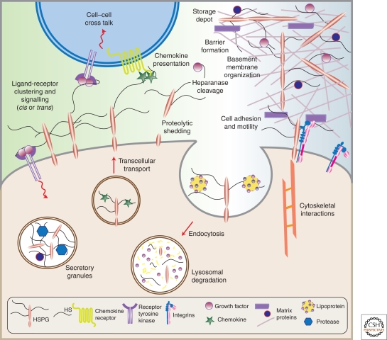
HSPGs have multiple activities in cells and tissues. (Adapted from Bishop et al. 2007; reprinted with permission from Nature Publishing Group © 2007.)
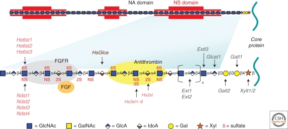
Heparan sulfate (HS) structure. HS biosynthesis initiates by the attachment of xylose to specific serine residues in HSPG core proteins followed by the formation of a linkage tetrasaccharide, glucuronic acid-galactose-galactose-xylose (GlcA-Gal-Gal-Xyl). Extl3 attaches the first N-acetyl-d -glucosamine (GlcNAc) residue and an enzyme complex composed of Ext1 and Ext2 alternately adds GlcA and GlcNAc to the nascent chain. The chains simultaneously undergo a series of processing reactions that begins by the removal of the acetyl groups from clusters of GlcNAc residues and substitution of the free amino groups with sulfate, catalyzed by one or more N-deacetylase-N-sulfotransferases (Ndst). The C5 epimerase (HsGlce) epimerizes d -glucuronic acids immediately adjacent to N-sulfoglucosamine units to l -iduronic acid (IdoA). A series of O-sulfotransferases can then add sulfate: uronyl 2-O-sulfotransferase (Hs2st) adds sulfate at C2 of the iduronic acids (and less frequently to glucuronic acids), 6-O-sulfotransferases (Hs6st1-3) add sulfate at C6 of the N-sulfoglucosamine units and less frequently to N-acetylglucosamine, and 3-O-sulfotransferases (Hs3st1, 2, 3a, 3b, 4, 5, 6) add sulfate at C3 of glucosamine units (N-sulfated or N-unsubstituted). As shown in the top of the figure by red shading, the modifications occur in clusters of variable length (N-sulfated or NS domains), which are interspersed by unmodified domains (N-acetylated or NA domains). The regions at the junction of these domains are sometimes called NA/NS domains (not shown) because the extent of processing is less. The modified domains make up binding sites for protein ligands as depicted for antithrombin, FGF and FGF receptor. The HS chains can be further modified once they arrive at the cell surface or in the ECM by two endosulfatases (Sulf1 and Sulf2), which remove specific sulfate groups located at C6 of glucosamine units, or by the action of extracellular heparanase or extracellular proteases (not shown). (Figure adapted from Bishop et al. 2007; reprinted with permission from Nature Publishing Group © 2007.)
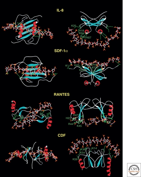
Docking of HS to chemokines. Molecular modeling was used to dock a fully sulfated heparin-like chain to several chemokines. The proteins are represented by ribbons except for the side chains of the basic amino acids directly involved in polysaccharide binding (green). The heparin molecule is represented by sticks. (Data from Lortat-Jacob et al. 2002.)
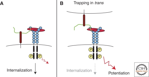
Model for trans-activation of VEGF receptor by HSPGs. (A) Resident plasma membrane HSPGs can mediate VEGF interactions with its receptor in cis, inducing cell signaling and subsequent internalization of the complex. (B) HSPGs from an adjacent cell can also mediate VEGF interactions with its receptor in trans, delaying internalization of the signaling complex and enhancing VEGF response. (From Jakobsson et al. 2006; reprinted with permission from Elsevier © 2006.)
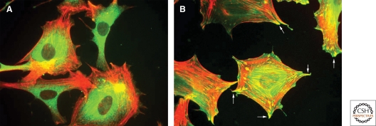
The role of syndecan-4 in focal adhesion. (A) Fibroblasts attach and spread through α5β1 integrin on coverslips coated with the integrin-binding domain of fibronectin but they do not form focal adhesions. (B) Focal adhesions (arrows) form only after engagement of syndecan-4 HS chains after the addition of the heparin-binding domain (HepII) from fibronectin. (Data for image from Okina et al. 2009.)
References
-
- Abrink M, Grujic M, Pejler G 2004. Serglycin is essential for maturation of mast cell secretory granule. J Biol Chem 279: 40897–40905 - PubMed
-
- Ai X, Kitazawa T, Do AT, Kusche-Gullberg M, Labosky PA, Emerson CP Jr 2007. SULF1 and SULF2 regulate heparan sulfate-mediated GDNF signaling for esophageal innervation. Development 134: 3327–3338 - PubMed
Publication types
MeSH terms
Substances
Grants and funding
LinkOut - more resources
Full Text Sources
Other Literature Sources
Medical
Molecular Biology Databases