. 2011 Aug;31(16):3196-207.
doi: 10.1128/MCB.05073-11. Epub 2011 Jun 20.

Posttranscriptional control of type I interferon genes by KSRP in the innate immune response against viral infection

Affiliations

Posttranscriptional control of type I interferon genes by KSRP in the innate immune response against viral infection

Wei-Jye Lin et al. Mol Cell Biol. 2011 Aug.

Abstract

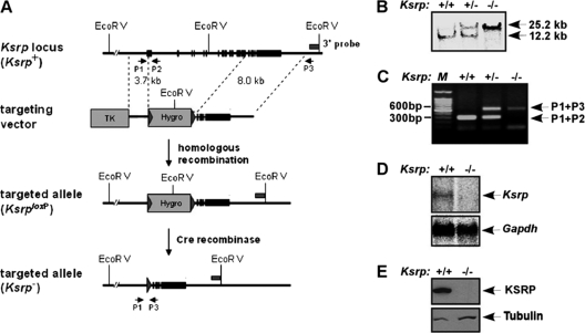
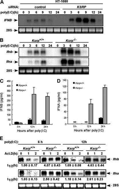
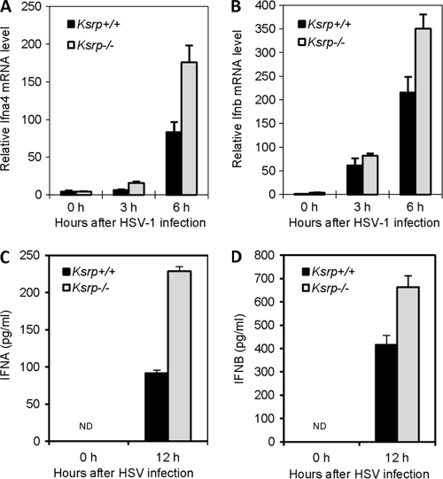
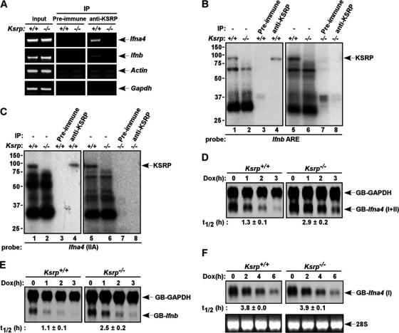
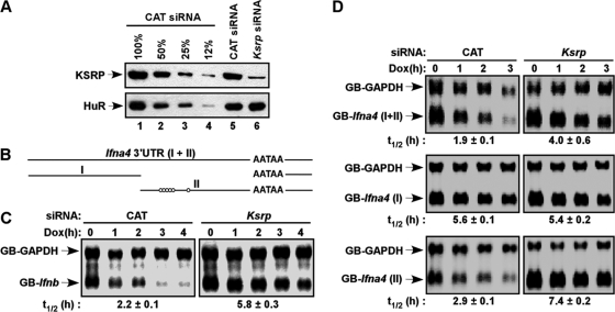
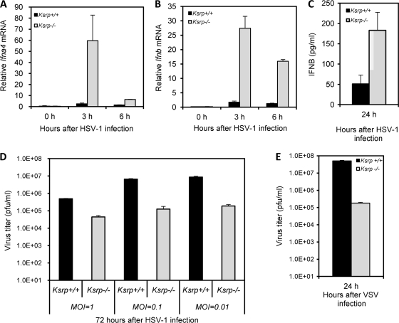
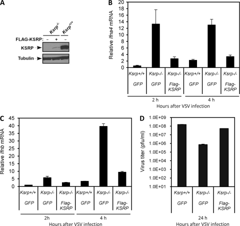
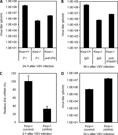
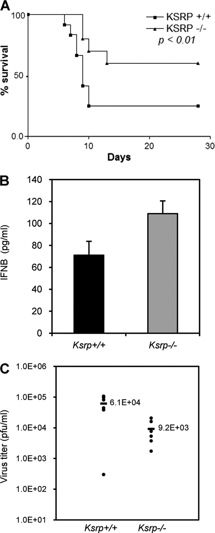
Inherently unstable mRNAs contain AU-rich elements (AREs) in the 3' untranslated regions. Expression of ARE-containing type I interferon transcripts is robustly induced upon viral infection and rapidly shut off thereafter. Their transient accumulation is partly mediated through posttranscriptional regulation. Here we show that mouse embryonic fibroblasts derived from knockout mice deficient in KH-type splicing regulatory protein (KSRP), an RNA-binding protein required for ARE-mediated mRNA decay, produce higher levels of Ifna and Ifnb mRNAs in response to viral infection as a result of decreased mRNA decay. Functional analysis showed that KSRP is required for the decay of Ifna4 and Ifnb mRNAs by interaction with AREs. The increased IFN expression renders Ksrp(-)(/)(-) cells refractory to herpes simplex virus type 1 and vesicular stomatitis virus infection. These findings support a role of a posttranscriptional mechanism in the control of type I IFN expression and highlight the function of KSRP in innate immunity by negatively regulating IFN production.


Figures

Fig. 1.
Fig. 1.
Generation of Ksrp knockout mice. (A) The wild-type Ksrp allele, the targeting vector, the targeted allele, and the genotyping methods are described. A hygromycin selection cassette with loxP sites flanked at both ends was designed to replace exon 1 to exon 13 of the mouse Ksrp allele. The hygromycin cassette in the targeted Ksrp allele was further excised by expression of Cre recombinase in ES cells. EcoRV sites, a 3′ external probe, and PCR primers used for genotyping are indicated. (B) Southern blot analysis shows that the Ksrp allele is targeted. Genomic DNA from the second generation of mouse offspring was digested with EcoRV and hybridized with 32P-labeled 3′ external probe. (C) Genotyping by PCR analysis. Genomic DNA was analyzed by PCR using P1, P2, and P3 primers to specifically amplify the wild-type (300-bp) and the targeted (600-bp) Ksrp alleles. (D) Northern blot analysis shows undetectable Ksrp mRNA expression in Ksrp-null mice. Poly(A)+ mRNA was isolated from wild-type and Ksrp/ livers and analyzed by using a 32P-labeled Ksrp cDNA probe. The blot was reprobed with a Gapdh probe. (E) Immunoblot analysis shows no detection of KSRP expression. Total protein was extracted from wild-type and Ksrp/ livers and subjected to immunoblotting using a monoclonal anti-KSRP antibody. The same blot was rehybridized with antibodies against α-tubulin.
Fig. 2.
Fig. 2.
Increased type I IFN gene expression in Ksrp/ MEFs. (A) Human fibrosarcoma HT-1080 cells were transfected with a control siRNA or a KSRP siRNA. At 48 h after transfection, cells were stimulated with poly(I·C) (10 μg/ml). Ifnb mRNA levels were analyzed by Northern blotting at different time points after poly(I·C) treatment. (B) Wild-type or Ksrp-deficient immortalized MEFs were treated with poly(I·C) (2 μg/ml). Total RNA was isolated at different time points and analyzed with probes against the coding region of Ifnb or Ifna4. The Ifna4 probe also recognizes other Ifna subtypes, due to their high homologies. (C and D) IFNB and IFNA levels in wild-type and Ksrp/ MEFs were analyzed by ELISA 12 h and 24 h after poly(I·C) treatment. Data are mean ± standard errors of the means of three experiments (IFNB, P < 0.01 at 12 h and P < 0.001 at 24 h; IFNA, P < 0.001 at 24 h). (E) Elevated IFN gene expression in Ksrp/ MEFs is due to increased mRNA stability. Wild-type or Ksrp-deficient MEFs were treated with poly(I·C) for 6 h or 12 h, and RNA was isolated at different time points after the addition of actinomycin D. mRNA levels of Ifna and Ifnb were analyzed by Northern blotting and quantitated with a phosphorimager. The calculated t1/2 values are shown as means ± standard deviations (n = 3).
Fig. 3.
Fig. 3.
Elevated IFN expression in primary Ksrp/ BMDCs. (A and B) BMDCs derived from Ksrp+/+ and Ksrp/ mice were infected with HSV-1 (MOI, 3), and total RNA was isolated at different time points. Quantitative RT-PCR was used to analyze the levels of Ifna4 (A) and Ifnb (B). Data are means ± standard errors of the means (n = 3; Ifna4, P < 0.01 at 3 h, P < 0.02 at 6 h; Ifnb, P < 0.02 at 6 h). (C and D) IFNA and IFNB levels in the supernatants of Ksrp+/+ and Ksrp/ BMDCs were analyzed by ELISA 12 h after HSV-1 infection. Data are means ± standard errors of the means of three experiments (IFNA, P < 0.001 at 12 h; IFNB, P < 0.01 at 12 h).
Fig. 4.
Fig. 4.
Regulation of Ifna4 and Ifnb mRNA stability by KSRP through interaction with the 3′-UTRs. (A) Association of Ifna4 and Ifnb transcripts with KSRP. Extracts of wild-type and Ksrp/ MEFs treated with poly(I·C) were subjected to immunoprecipitation with preimmune serum or anti-KSRP serum. The coprecipitated RNA was isolated, and the presence of Ifna4 and Ifnb transcripts was analyzed by RT-PCR. The presence of Ifna4 and Ifnb transcripts in the input is also shown. The presence of β-actin and Gapdh mRNAs was also analyzed as controls for specificity. (B and C) KSRP interacts with the AREs of Ifnb and Ifna4. Wild-type or Ksrp/ extracts were incubated with 32P-labeled RNAs composed of the AREs of Ifnb (B) or Ifna4 (C) (sequence available on request) and subjected to UV cross-linking. The RNA-protein complexes were either directly analyzed by SDS-PAGE (lanes 1, 2, 5, and 6) or subjected to immunoprecipitation with preimmune serum (lanes 3 and 7) or anti-KSRP serum (lanes 4 and 8) and then the immunoprecipitates were analyzed by SDS-PAGE and autoradiography. Note some variations of protein-binding levels to Ifna4 ARE in different experiments. (D and E) The 3′-UTRs of Ifna4 and Ifnb confer mRNA instability and regulation by KSRP. Wild-type and Ksrp/ MEFs were cotransfected with a construct expressing β-globin mRNA reporter containing either the 3′-UTR of Ifna4, GB-Ifna4(I+II) (D) or the 3′-UTR of Ifnb, GB-Ifnb (E) under the control of a tetracycline-responsive promoter, a plasmid expressing a β-globin reporter consisting of the 3′-UTR of Gapdh under the control of the cytomegalovirus promoter (GB-Gapdh), and a plasmid expressing tetracycline-responsive transactivator (tTA). At 6 h after transcriptional induction, doxycycline (2 μg/ml) was added to block transcription. Total RNA was isolated at different time points. The levels of GB-Ifna4(I+II), GB-Ifnb, and GB-Gapdh mRNAs were analyzed by Northern blotting and quantitated. The calculated half-lives of GB-Ifna4(I+II) and GB-Ifnb mRNAs are shown as means ± standard deviations (n = 3). (F) The decay of a globin mRNA containing the non-ARE region of Ifna4, GB-Ifna4(I), was analyzed in Ksrp+/+ or Ksrp/ MEFs as described for panel D. The calculated half-lives of GB-Ifna4(I) mRNA are shown as means ± standard deviations (n = 3).
Fig. 5.
Fig. 5.
Decay of mRNA reporters consisting of the 3′-UTRs was impaired in KSRP-downregulated NIH 3T3 cells. (A) Immunoblot analysis showed downregulation of KSRP in NIH 3T3-B2A2 cells transfected with a KSRP siRNA. Different amounts of extracts from cells transfected with a control (chloramphenicol [CAT]) siRNA were loaded in lanes 1 to 4 to show the efficiency of knockdown. The levels of KSRP and HuR were analyzed by immunoblotting. (B) Schematic diagram showing the 3′-UTR of Ifna4, which is further divided into two subregions (I and II). Open circles indicate AUUUA motifs. (C and D) NIH 3T3-B2A2 cells, stably expressing a tetracycline-responsive transactivator, were transfected with constructs expressing globin reporters composed of the 3′-UTR of Ifnb (C) or different regions of the Ifna4 3′-UTR (D) under the control of a Tet regulatory promoter and a construct constitutively expressing GB-Gapdh mRNA under the control of the cytomegalovirus promoter. Total RNA was isolated at different time points after addition of doxycycline (Dox). The levels of reporter mRNAs were analyzed by Northern blotting. The calculated half-lives (t1/2) of reporter mRNAs are shown (n = 3).
Fig. 6.
Fig. 6.
Replication levels of HSV-1 and VSV are markedly reduced in Ksrp/ MEFs. (A and B) Ksrp+/+ and Ksrp/ MEFs were infected with HSV-1 (MOI, 3). The levels of Ifna4 (A) and Ifnb (B) were analyzed by qRT-PCR. Values represent means ± standard errors of the means (n = 3; Ifna4, P < 0.05 at 3 h and P < 0.001 at 6 h; Ifnb, P < 0.005 at 3 h and P < 0.001 at 6 h). (C) IFNB levels in culture supernatants of Ksrp+/+ and Ksrp/ MEFs were analyzed by ELISA 24 h after HSV-1 infection. Data are means ± standard errors of the means (n = 3; P < 0.03). (D) Reduced HSV-1 replication in Ksrp/ MEFs. Ksrp+/+ and Ksrp/ MEFs were infected with HSV-1 using different MOIs (1, 0.1, or 0.01). At 72 h after infection, viral production was analyzed by plaque assay. Data are means ± standard errors of the means of three independent experiments (MOI, 1 [P < 0.0001]; MOI, 0.1 [P < 0.0002]; MOI, 0.01 [P < 0.001]). (E) Reduced VSV replication in Ksrp/ MEFs. Ksrp+/+ and Ksrp/ MEFs were infected with VSV at an MOI of 0.2. At 24 h after infection, viral production was analyzed by plaque assay. Data are means ± standard errors of the means of three experiments (P < 0.004).
Fig. 7.
Fig. 7.
The viral resistance of Ksrp/ MEFs is associated with a lack of KSRP. (A) Reexpression of KSRP in Ksrp/ MEFs. Protein samples from control retrovirus (GFP)-transduced Ksrp+/+ and Ksrp/ MEFs, or KSRP-expressing retrovirus-transduced Ksrp/ MEFs, were analyzed by immunoblotting using anti-KSRP antibody. The blot was rehybridized with anti-α-tubulin antibody. (B and C) Decreased IFN mRNA expression in Ksrp/ MEFs reexpressing KSRP. MEFs were infected with VSV (MOI, 1). The mRNA levels of Ifna4 (B) and Ifnb (C) were analyzed by qRT-PCR 2 h and 4 h after infection. Values represent means ± standard errors of the means (n = 3). (D) Increased VSV replication in Ksrp/ MEFs reexpressing KSRP. MEFs were infected with VSV (MOI, 1). VSV production was analyzed 24 h after infection by plaque assay. Data are means ± standard errors of the means of three experiments.
Fig. 8.
Fig. 8.
Enhanced IFN expression is responsible for the viral resistance in Ksrp/ MEFs. (A and B) Ksrp/ MEFs were infected with VSV (MOI, 0.01) and incubated with neutralizing antibodies against both IFNA and IFNB (A) or against IFNAR1 (B) or control IgG. VSV production was analyzed 24 to 36 h after infection by plaque assay. Data are means ± standard errors of the means of three experiments (Ksrp/ [PI] versus Ksrp/ [anti-IFN], P < 0.001; Ksrp/ [IgG] versus Ksrp/ [IFNAR1], P < 0.001). (C and D) Ksrp/ MEFs were infected with lentiviral shRNAs against Ifnb or shRNA against GFP. Transduced cells were infected with VSV (MOI, 1). Expression of Ifnb was analyzed by qRT-PCR 2 h postinfection, and the virus titer was analyzed 24 h after infection. Data are means ± standard errors of the means of three experiments (P < 0.005).
Fig. 9.
Fig. 9.
Ksrp/ mice are resistant to HSV-1 infection. (A) Ksrp+/+ and Ksrp/ mice were intracranially infected with 300 PFU of HSV-1. The survival of the infected mice was monitored for 28 days. Kaplan-Meier survival curves are shown (animals surviving at the end of experiment: Ksrp+/+, 5/17; Ksrp/, 11/16; P < 0.01, log-rank test). The median survival was 9 days for Ksrp+/+ mice and >28 days for Ksrp/ mice. (B) HSV-1-infected Ksrp+/+ and Ksrp/ brains were harvested 3 days after infection. IFNB levels were analyzed by ELISA. Values are means ± standard errors of the means (n = 3; P < 0.05). (C) HSV-1-infected Ksrp+/+ and Ksrp/ brains were harvested 6 days after infection. Virus production was analyzed by plaque assay (Ksrp+/+, n = 6; Ksrp/, n = 6; P < 0.02).

References

    1. Asselin-Paturel C., et al. 2001. Mouse type I IFN-producing cells are immature APCs with plasmacytoid morphology. Nat. Immunol. 2:1144–1150 - PubMed
    1. Asselin-Paturel C., Trinchieri G. 2005. Production of type I interferons: plasmacytoid dendritic cells and beyond. J. Exp. Med. 202:461–465 - PMC - PubMed
    1. Bakheet T., Williams B. R., Khabar K. S. 2003. ARED 2.0: an update of AU-rich element mRNA database. Nucleic Acids Res. 31:421–423 - PMC - PubMed
    1. Bakheet T., Williams B. R., Khabar K. S. 2006. ARED 3.0: the large and diverse AU-rich transcriptome. Nucleic Acids Res. 34:D111–D114 - PMC - PubMed
    1. Banchereau J., Pascual V. 2006. Type I interferon in systemic lupus erythematosus and other autoimmune diseases. Immunity 25:383–392 - PubMed

Publication types

LinkOut - more resources