. 2011 Jun 27;193(7):1213-28.
doi: 10.1083/jcb.201103076. Epub 2011 Jun 20.

Cdc55 coordinates spindle assembly and chromosome disjunction during meiosis

Affiliations

Cdc55 coordinates spindle assembly and chromosome disjunction during meiosis

Farid Bizzari et al. J Cell Biol. .

Abstract

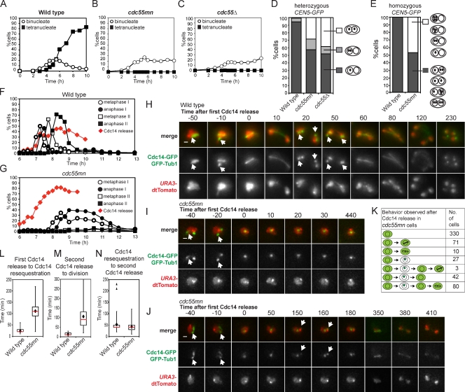
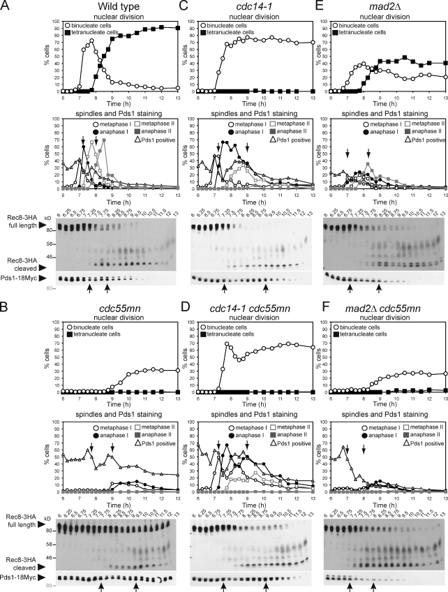
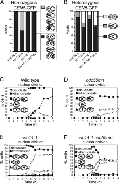
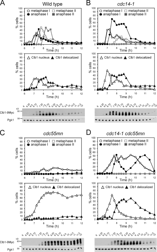
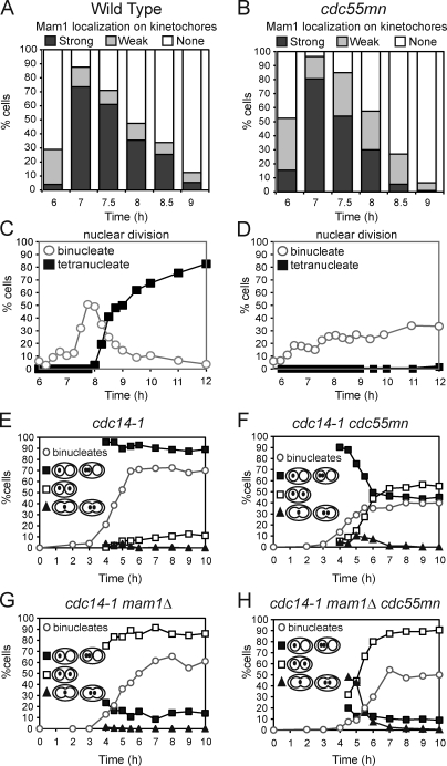
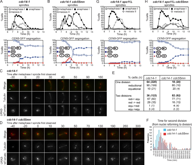
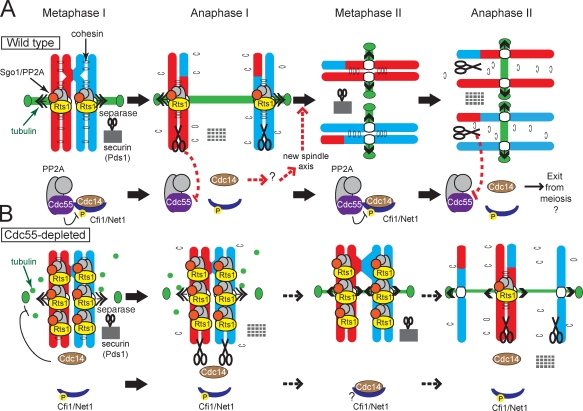
During meiosis, two consecutive nuclear divisions follow a single round of deoxyribonucleic acid replication. In meiosis I, homologues are segregated, whereas in meiosis II, sister chromatids are segregated. This requires that the sequential assembly and dissolution of specialized chromosomal factors are coordinated with two rounds of spindle assembly and disassembly. How these events are coupled is unknown. In this paper, we show, in budding yeast, that the protein phosphatase 2A regulatory subunit Cdc55 couples the loss of linkages between chromosomes with nuclear division by restraining two other phosphatases, Cdc14 and PP2A(Rts1). Cdc55 maintains Cdc14 sequestration in the nucleolus during early meiosis, and this is essential for the assembly of the meiosis I spindle but not for chromosomes to separate. Cdc55 also limits the formation of PP2A holocomplexes containing the alternative regulatory subunit Rts1, which is crucial for the timely dissolution of sister chromatid cohesion. Therefore, Cdc55 orders passage through the meiotic divisions by ensuring a balance of phosphatases.


Figures

Figure 1.
Figure 1.
Impaired nuclear division, random chromosome segregation, and ectopic Cdc14 release in cdc55mn cells. (A–D) Meiosis was induced in strains carrying heterozygous CEN5-GFP, PDS1-18MYC, and otherwise wild type (AM4796), cdc55mn (AM4891), or cdc55Δ (AM5338). The percentages of binucleate and tetranucleate cells (n = 200; A–C) or the pattern of GFP foci in binucleate cells (n > 800; D) is shown for a representative experiment. (E) Wild-type (AM6040) and cdc55mn (AM5936) strains carrying homozygous CEN5-GFP were analyzed as described in D. (F and G) Wild-type (AM6633) and cdc55mn (AM6626) cells carrying 3HA-CDC14, GAL-NDT80, and pGPD1-GAL4(848).ER were induced to sporulate and released from a pachytene block at 6 h. The percentages of cells with the indicated spindle morphology and with Cdc14 released from the nucleolus are shown for a representative experiment. (H–K) Wild-type (AM6935) and cdc55mn (AM6942) cells carrying CDC14-GFP, GFP-TUB1, and homozygous tetR-tdTomato were filmed. (H–J) Still images from Videos 1 (wild-type; H), 2 (cdc55mn; I), and 3 (cdc55mn; J). Arrows indicate Cdc14 sequestered in the nucleolus. Bars, 1 µm. (K) Behavior of cdc55mn cells that were in prophase I (as judged by spindle morphology) at the start of filming and released Cdc14 from the nucleolus (563/1,203 prophase I cells). Examples of extruding microtubules are shown in fixed cells in Fig. S2. Times are given in minutes. (L–N) The time elapsed between the first Cdc14 release to Cdc14 resequestration (L), the second Cdc14 release to nuclear division (M), or Cdc14 resequestration and rerelease (N) are shown for cells that were in prophase I at the start of filming and in which two rounds of Cdc14 release were observed (wild type, n = 95; cdc55mn, n = 80). Box boundaries represent the bottom quartile and top quartile. The red line indicates the median, diamonds indicate the mean, and error bars represent the minimum and maximum values observed except for in the case of N in which two outliers (triangles) were excluded from the analysis. Because images were captured at 10-min intervals, the time elapsed between two observed events is subject to an error of 20 min.
Figure 2.
Figure 2.
Impaired spindle assembly in cdc55mn cells is caused by ectopic Cdc14 activation. (A–F) Wild-type (AM6142; A), cdc55mn (AM6131; B), cdc14-1 (AM7549; C), cdc14-1 cdc55mn (AM7550; D), mad2Δ (AM7547; E), and mad2Δ cdc55mn (AM7548; F) strains carrying REC8-3HA, PDS1-18MYC, GAL-NDT80, and pGPD1-GAL4(848).ER were cultured as described in Fig 1 (F and G). The percentages of binucleate and tetranucleate cells, Pds1-positive cells, and cells with the indicated spindle morphology were determined. Anti-HA and anti-Myc immunoblots showing the positions of full-length Rec8-3HA, cleaved Rec8-3HA, and Pds1-18Myc (arrowheads) are shown with protein molecular mass markers in black (anti-HA) or gray (anti-Myc). Arrows indicate a reduction in Pds1-positive cells in the first and second meiotic division. Blots are representative (A and B) or were performed once (C–F). Numbers at the top of the blots indicate time in hours.
Figure 3.
Figure 3.
The cdc14-1 mutation rescues homologue segregation in cdc55mn cells but does not prevent equational segregation on the same axis. (A) Segregation of homozygous CEN5-GFP foci in ≥1,000 binucleate cells of wild-type (AM6040), cdc55mn (AM5936), cdc14-1 (AM6902), and cdc14-1 cdc55mn (AM6908) strains were determined as described in Fig. 1 D. (B–F) Segregation of heterozygous CEN5-GFP in wild-type (AM4796), cdc55mn (AM4891), cdc14-1 (AM6910), and cdc14-1 cdc55mn (AM6934) cells. (B) Segregation in binucleate cells was determined as described in A. (C–F) The total percentage of binucleate and tetranucleate cells and the percentage of binucleate cells that have GFP foci in one, both, or between the two nuclei are shown for a representative experiment.
Figure 4.
Figure 4.
Ectopic Cdc14 activation does not cause premature degradation of the meiotic cyclin Clb1. (A–D) Wild-type (AM6770; A), cdc14-1 (AM7815; B), cdc55mn (AM6961; C), and cdc14-1 cdc55mn (AM7816; D) cells carrying CLB1-9MYC, GAL-NDT80, and pGPD1-GAL4(848).ER were released from a pachytene arrest. The percentages of cells with the indicated spindle morphology (top) or Clb1-9Myc localization (middle) were determined at the indicated time points. The anti-Myc immunoblot (bottom) is shown with the anti-Pgk1 immunoblot as a loading control and the positions of molecular mass markers indicated. Western blots are representative (A and C) or were performed once (B and D). Numbers at the top of the blots indicate time in hours.
Figure 5.
Figure 5.
Kinetochores are monooriented during meiosis I in cdc55mn cells. (A–D) Wild-type (AM5900; A and C) and cdc55mn (AM5771; B and D) cells carrying MAM1-9MYC, NDC10-6HA, GAL-NDT80, and pGPD1-GAL4(848).ER were released from a pachytene block, and the localization of Mam1-9Myc was determined using Ndc10-6HA as a marker for kinetochores. (C and D) The percentages of binucleate and tetranucleate cells determined at the indicated time points are shown for a representative experiment. (E–H) Strains carrying heterozygous CEN5-GFP dots and otherwise cdc14-1 (AM6910), cdc14-1 cdc55mn (AM6934), cdc14-1 mam1Δ (AM7355), and cdc14-1 mam1Δ cdc55mn (AM7328) were analyzed as described in Fig. 3 (C–F).
Figure 6.
Figure 6.
Equational segregation occurs on a newly assembled spindle in cdc14-1 mutants and is elevated by Cdc55 depletion. (A and B) Strains carrying heterozygous CEN5-GFP, GAL-NDT80, pGPD1-GAL4(848).ER, and either cdc14-1 (AM8044; A) or cdc14-1 cdc55mn (AM8045; B) were released from a pachytene block, and the percentages of cells with the indicated spindle morphology (top graph) and pattern of CEN5-GFP localization in the binucleate cells (bottom graph) were determined. (C–F) cdc14-1 (AM7866) and cdc14-1 cdc55mn (AM7867) cells carrying URA3-tdTomato (red) and GFP-TUB1 (green) were imaged at 10-min intervals for a total of 9 h and 46 min and 9 h and 56 min, respectively. (C) Example of a cdc14-1 cell that completes a single meiotic division. (D) Example of a cdc14-1 cdc55mn cell that completes two divisions on the same axis. Times at the top are given in minutes. Bars, 1 µm. (E) Behavior of cdc14-1 and cdc14-1 cdc55mn cells that were initially in prophase I and which performed at least one division during the course of filming. red, reductional; equ, equational. (F) The frequency of cdc14-1 and cdc14-1 cdc55mn cells that performed the second division within the indicated time period. Also see Videos 4 and 5. (G and H) Strains carrying heterozygous CEN5-GFP dots GAL-NDT80, pGPD1-GAL4(848).ER, and either spo11Δ cdc14-1 (AM8046; G) or spo11Δ cdc14-1 cdc55mn (AM8047; H) were treated as described in A and B.
Figure 7.
Figure 7.
Overprotection of cohesin in a cdc55mn cell during meiosis. (A–F) Wild-type (AM7228; A, C, and E) and cdc55mn (AM7229; B, D, and F) cells carrying GAL-NDT80 and pGPD1-GAL4(848).ER were released from the pachytene block, and Sgo1 localization at kinetochores (A and B) or the percentages of binucleate and tetranucleate cells (C and D) were determined in a single experiment. (E and F) Examples of Sgo1 localization are shown. Bars, 2 µm. (G and H) Depletion of Sgo1 (G) or replacement of Rec8 by Scc1 (H) abolishes equational segregation in cdc14-1 cdc55mn cells. Strains carrying heterozygous CEN5-GFP and with the indicated genotypes were treated as described in Fig. 1. n = 680–2,000. Strains used were wild type (AM4796), cdc55mn (AM4891), sgo1mn (AM4911), sgo1mn cdc55mn (AM7286), cdc14-1 (AM6910), cdc14-1 cdc55mn (AM6934), cdc14-1 sgo1mn (AM7360), cdc14-1 sgo1mn cdc55mn (AM7421), spo11Δ rec8Δ pREC8-SCC1 (AM5501), spo11Δ rec8Δ pREC8-SCC1 cdc55mn (AM5502), spo11Δ rec8Δ pREC8-SCC1 cdc14-1 (AM7361), and spo11Δ rec8Δ pREC8-SCC1 cdc14-1 cdc55mn (AM7362). (I) Tpd3-6HA was immunoprecipitated using anti-HA antibodies from meiotic extracts of wild-type and cdc55mn cells carrying RTS1-3PK and either TPD3-6HA (AM8028 and AM8014) or no tag (AM8012 and AM8029). Anti-V5 (PK) and anti-HA immunoblots of input and immunoprecipitated samples from strains of the indicated genotypes are shown, with protein molecular mass markers shown in black or red, respectively. An Rts1-3PK degradation product is indicated by the gray arrowhead, and the asterisk indicates residual 3HA-Cdc55. (J) qPCR analysis of chromatin immunoprecipitated using anti-V5 (PK) antibodies from cdc20mn (AM3560), cdc20mn RTS1-3PK (AM7902), cdc20mn cdc55mn (AM7903), and cdc20mn cdc55mn RTS1-3PK (AM7904) strains. The mean of three experiments is shown with error bars indicating standard deviation.
Figure 8.
Figure 8.
Model for coordination of the meiotic program by Cdc55. (A) In wild-type cells during metaphase I, PP2ACdc55 both maintains Cdc14 sequestration in the nucleolus by dephosphorylating Cfi1 and restricts the amount of PP2A scaffold (A) and catalytic (C) subunits available to form a complex with Rts1 (B′). The level of PP2ARts1 defines the domain of cohesion that will be protected during meiosis I. Once chromosomes are correctly aligned, separase activation leads to both the cleavage of unprotected arm cohesin and, through PP2ACdc55 down-regulation, Cdc14 release. Cdc14 release during anaphase I ensures that a second spindle axis will assemble in meiosis II after its resequestration. (B) In cdc55mn cells, ectopic Cdc14 release interferes with spindle assembly, and excess PP2ARts1 extends the domain of protected cohesin. This prevents nuclear division except in a low number of cells in which Cdc14 is relocalized to the nucleolus through an unknown mechanism (?). P indicates Cdk-dependent phosphorylation of Cfi1/Net1.

References

    1. Azzam R., Chen S.L., Shou W., Mah A.S., Alexandru G., Nasmyth K., Annan R.S., Carr S.A., Deshaies R.J. 2004. Phosphorylation by cyclin B-Cdk underlies release of mitotic exit activator Cdc14 from the nucleolus. Science. 305:516–519 10.1126/science.1099402 - DOI - PubMed
    1. Bajpai R., Makhijani K., Rao P.R., Shashidhara L.S. 2004. Drosophila Twins regulates Armadillo levels in response to Wg/Wnt signal. Development. 131:1007–1016 10.1242/dev.00980 - DOI - PubMed
    1. Benjamin K.R., Zhang C., Shokat K.M., Herskowitz I. 2003. Control of landmark events in meiosis by the CDK Cdc28 and the meiosis-specific kinase Ime2. Genes Dev. 17:1524–1539 10.1101/gad.1101503 - DOI - PMC - PubMed
    1. Breitkreutz A., Choi H., Sharom J.R., Boucher L., Neduva V., Larsen B., Lin Z.Y., Breitkreutz B.J., Stark C., Liu G., et al. 2010. A global protein kinase and phosphatase interaction network in yeast. Science. 328:1043–1046 10.1126/science.1176495 - DOI - PMC - PubMed
    1. Buonomo S.B., Rabitsch K.P., Fuchs J., Gruber S., Sullivan M., Uhlmann F., Petronczki M., Tóth A., Nasmyth K. 2003. Division of the nucleolus and its release of CDC14 during anaphase of meiosis I depends on separase, SPO12, and SLK19. Dev. Cell. 4:727–739 10.1016/S1534-5807(03)00129-1 - DOI - PubMed

Publication types

MeSH terms