. 2011 Jul 5;108(27):11246-51.
doi: 10.1073/pnas.1012401108. Epub 2011 Jun 20.

Neurotrophin-mediated dendrite-to-nucleus signaling revealed by microfluidic compartmentalization of dendrites

Affiliations

Neurotrophin-mediated dendrite-to-nucleus signaling revealed by microfluidic compartmentalization of dendrites

Michael S Cohen et al. Proc Natl Acad Sci U S A. .

Abstract

Signaling from dendritic synapses to the nucleus regulates important aspects of neuronal function, including synaptic plasticity. The neurotrophin brain-derived neurotrophic factor (BDNF) can induce long-lasting strengthening of synapses in vivo and this effect is dependent on transcription. However, the mechanism of signaling to the nucleus is not well understood. Here we describe a microfluidic culture device to investigate dendrite-to-nucleus signaling. Using these microfluidic devices, we demonstrate that BDNF can act directly on dendrites to elicit an anterograde signal that induces transcription of the immediate early genes, Arc and c-Fos. Induction of Arc is dependent on dendrite- and cell body-derived calcium, whereas induction of c-Fos is calcium-independent. In contrast to retrograde neurotrophin-mediated axon-to-nucleus signaling, which is MEK5-dependent, BDNF-mediated anterograde dendrite-to-nucleus signaling is dependent on MEK1/2. Intriguingly, the activity of TrkB, the BDNF receptor, is required in the cell body for the induction of Arc and c-Fos mediated by dendritically applied BDNF. These results are consistent with the involvement of a signaling endosome-like pathway that conveys BDNF signals from the dendrite to the nucleus.


Conflict of interest statement

Conflict of interest statement: N.L.J. is an inventor and patent holder of the microfluidic devices described in this article and is a cofounder of Xona Microfluidics LLC, which markets related microfluidic devices.

Figures

Fig. 1.
Fig. 1.
A microfluidic device to fluidically isolate dendrites from the cell body. (A) Schematic representation of the neuronal microfluidic device. The device is fabricated from a PDMS mold containing mirror image compartments (1.5-mm wide, 7-mm long, 100-μm high) connected by microgrooves (10-μm wide, 3-μm high). (B) Phase-contrast image of the microfluidic device with 75-μm long microgrooves illustrating schematic representations of CNS neurons projecting dendrites and axons into the neurite compartment. (CE) Immunofluorescence analysis of E18 rat cortical neurons (DIV14) cultured in the microfluidic device. (C) Extensive crossing of dendrites (MAP2, green) into the neurite compartment is observed for cell bodies within ∼100 μm of the microgrooves. Cell bodies (DAPI, blue) are restricted to the cell body compartment. (Scale bar, 50 μm.) (D) Axons (TAU-1, red) project extensively into the neurite compartment and considerably farther from the microgrooves than the dendrites (MAP2, green), which remain close to the microgrooves. (Scale bar, 20 μm.) (E) A cortical neuron expressing GFP (green) reveals the presence of dendritic spines on dendrites (MAP2, red) in the neurite compartment. (Scale bar, 20 μm.) (Inset) Higher magnification image of a segment of dendrite enclosed by the white box in the merged image. (Scale bar, 2 μm.)
Fig. 2.
Fig. 2.
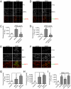
BDNF acts at dendrites to induce the expression Arc and c-Fos. On DIV10 BDNF (100 ng/mL) or vehicle control was applied to the neurite (A and B) or the cell body (E and F) compartment. After 2 h, cells were fixed and immunolabeled with antibodies against Arc (green, A and E), c-Fos (green, B and F), and MAP2 (red). (Scale bars, 20 μm.) (A and B) The cell bodies in the cell body compartment that have dendrites projecting into the neurite compartment are indicated by white arrowheads. (C and D) Induction of Arc and c-Fos expression upon dendritic BDNF stimulation is because of new transcription. On DIV10, actinomycin D (2 μg/mL) or vehicle control was added to the cell body compartment for 1 h followed by the addition of BDNF (100 ng/mL) to the neurite compartment for 2 h. Cells were fixed and immunolabeled with antibodies against c-Fos and MAP2. (G and H) Quantification of results from A, B, E, and F. (I) Local protein synthesis is not required for c-Fos induction mediated by dendritic BDNF stimulation. On DIV10, CHX (10 μM) or vehicle control was added to the indicated compartment for 1 h followed by the addition of BDNF (100 ng/mL) to the neurite compartment for 2 h. Cells were fixed and immunolabeled with antibodies against c-Fos and MAP2. The error bars represent SEM, ***P < 0.0001, **P = 0.006 (unpaired, two-tailed t test); n values listed above the bars represent the number of cell bodies analyzed.
Fig. 3.
Fig. 3.
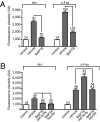
Induction of Arc by dendritically-applied BDNF is dependent on dendrite- and cell body-derived calcium, whereas induction of c-Fos is calcium-independent. (A) On DIV10, BAPTA-AM (32 μM) or vehicle control was added to the cell body compartment followed by the addition of BDNF (100 ng/mL) to the cell body (A) or neurite (B) compartment for 2 h. Cells were fixed and immunolabeled with antibodies against Arc, c-Fos, and MAP2. The error bars represent SEM, ***P < 0.0001 (unpaired, two-tailed t test); n values listed above the bars represent the number of cell bodies analyzed.
Fig. 4.
Fig. 4.
Trk and Erk1/2 activity in the cell body are required for the induction of Arc and c-Fos mediated by dendritically applied BDNF. (A and B) On DIV10, K252a (1 μM) or vehicle control was added to the indicated compartment followed by the addition of BDNF (100 ng/mL) to the neurite compartment for 2 h. Cells were fixed and immunolabeled with antibodies against Arc (A), c-Fos (B), and MAP2. (C and D) On DIV10, PD 0325901 (150 nM) or BI D1870 (10 μM) or vehicle control was added to the cell body compartment followed by the addition of BDNF (100 ng/mL) to the neurite compartment for 2 h. Cells were fixed and immunolabeled with antibodies against Arc (A), c-Fos (B), and MAP2. The error bars represent SEM, ***P < 0.0001, *P < 0.05 (unpaired, two-tailed t test); n values listed above the bars represent the number of cell bodies analyzed.

References

    1. Minichiello L. TrkB signalling pathways in LTP and learning. Nat Rev Neurosci. 2009;10:850–860. - PubMed
    1. Kang H, Welcher AA, Shelton D, Schuman EM. Neurotrophins and time: Different roles for TrkB signaling in hippocampal long-term potentiation. Neuron. 1997;19:653–664. - PubMed
    1. Korte M, et al. Hippocampal long-term potentiation is impaired in mice lacking brain-derived neurotrophic factor. Proc Natl Acad Sci USA. 1995;92:8856–8860. - PMC - PubMed
    1. Minichiello L, et al. Essential role for TrkB receptors in hippocampus-mediated learning. Neuron. 1999;24:401–414. - PubMed
    1. Patterson SL, et al. Recombinant BDNF rescues deficits in basal synaptic transmission and hippocampal LTP in BDNF knockout mice. Neuron. 1996;16:1137–1145. - PubMed

Publication types

MeSH terms

Substances

LinkOut - more resources