. 2011 Jul 5;108(27):11145-50.
doi: 10.1073/pnas.1015302108. Epub 2011 Jun 20.

A dynamical model of ommatidial crystal formation

Affiliations

A dynamical model of ommatidial crystal formation

David K Lubensky et al. Proc Natl Acad Sci U S A. .

Abstract

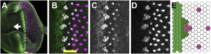
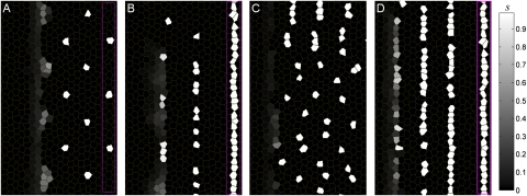
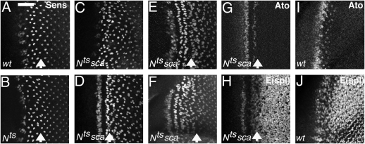
The crystalline photoreceptor lattice in the Drosophila eye is a paradigm for pattern formation during development. During eye development, activation of proneural genes at a moving front adds new columns to a regular lattice of R8 photoreceptors. We present a mathematical model of the governing activator-inhibitor system, which indicates that the dynamics of positive induction play a central role in the selection of certain cells as R8s. The "switch and template" patterning mechanism we observe is mathematically very different from the well-known Turing instability. Unlike a standard lateral inhibition model, our picture implies that R8s are defined before the appearance of the complete group of proneural cells. The model reproduces the full time course of proneural gene expression and accounts for specific features of the refinement of proneural groups that had resisted explanation. It moreover predicts that perturbing the normal template can lead to eyes containing stripes of R8 cells. We observed these stripes experimentally after manipulation of the Notch and scabrous genes. Our results suggest an alternative to the generally assumed mode of operation for lateral inhibition during development; more generally, they hint at a broader role for bistable switches in the initial establishment of patterns as well as in their maintenance.


Conflict of interest statement

The authors declare no conflict of interest.

Figures

Fig. 1.
Fig. 1.
Specification of R8 cells. In all figures, anterior is to the left. (A) In the eye imaginal disc epithelium, precisely constructed ommatidia each differentiate around a single R8 cell. R8 specification is accompanied by a transient contraction of disc cells in the “morphogenetic furrow” (MF) that sweeps anteriorly across the disc (arrow gives MF direction). Posterior to the furrow are vertical columns of R8s. Ato protein (green) appears just ahead of the furrow and is accompanied, then replaced, by Sens (magenta), expressed stably in R8 cells. (B) Ato expression evolves from a low uniform stripe into periodic intermediate groups (IGs) that then resolve into single R8 cells. Sens tracks Ato from the IGs. [Because the eye disc epithelium is pseudostratified, there are more unstained cells between R8s than it might appear from the size of the stained R8 nuclei (2, 4).] (C) Ato expression in intermediate groups and R8 cells. (Ato is lost in R8s posterior to the MF through a regulatory mechanism not discussed in this paper; ref. 33). (D) Sens expression. (E) Schematic of Ato (green) and Sens (magenta rings) expression in the furrow. Black dotted lines outline an IG, which has higher Ato expression than the stripe to its anterior. Yellow scale bars indicate the extent of the MF. (Scale bars: A and B, 18 μm.)
Fig. 2.
Fig. 2.
R8 determination network. (A) Interactions regulating the expression of Ato in the MF. Pointed arrows, activation; blunt arrows, inhibition. Green ellipses, nonautonomous signals; blue boxes, transcription factors acting cell autonomously. EGFR is known to block Hh signals (18); Dl and Sca are shown acting directly on Ato because it is not certain which mode of ato activation they inhibit. Pointed (Pnt) is activated in the photoreceptor differentiation pathway that eventually leads to hh transcription posterior to the MF (46). (B) Simplified mathematical model of the interactions in A. All inhibitory signals are condensed into the variable u, and all long-range activation into h. (C) Simulated a expression in the model; compare Fig. 1C. (Parameters are in Table S1.) (D) The patterning mechanism in the model. Each R8 (dots) inhibits a within a certain radius (circles). As the MF progresses, the first cell reached by the activator h that is free of this inhibition (blue) is strongly favored to become the next R8. a expression extends outward from this cell to transiently form the IG (green triangles).
Fig. 3.
Fig. 3.
R8 model patterns depend on initial conditions. Sens (s) expression in the model is shown. (See Fig. S6 for other model variables.) (A) Wild-type parameters (Table S1) with an ordered template yield the usual hexagonal pattern. (B) With a stripe-like template, the same wild-type parameter set generates poorly ordered isolated and twinned R8s, as seen after a perturbation of Notch function (Fig. 4B). (C) With a template of single R8s, the slower u (“sca”) parameters (Table S1) lead to a disrupted pattern with some twinned R8s (compare Fig. 4C). (D) With a stripe template, the sca parameter set maintains stripe-like R8 patterning. Such patterns have now been observed experimentally (Fig. 4D). Magenta boxes in A, B, and D enclose the initial template (Fig. S4B); the template in C is the same as in A but is omitted to leave more space for the full sca pattern.
Fig. 4.
Fig. 4.
R8 patterns after experimental perturbations. (AF) Projections of confocal images of Sens labeling. (A–H) Ommatidia near the arrow were at the IG stage at the higher (restrictive) temperature. (A) Normal patterning in wild type, exposed to 31.5 °C for 2 h and 18 °C for 14 h. Scale bar gives MF extent at fixation. (Scale bar, 17 μm.) (B) Nts (temperature-sensitive allele) exposed to the restrictive temperature of 31.5 °C for 2 h and returned to the permissive 18 °C for 14 h before fixation. R8 patterning is haphazard anterior to the arrow indicating ommatidia that were IGs at the restrictive temperature. (C) Disordered ommatidial development and sporadic R8 twinning in the sca mutant phenotype (Nts; sca at the permissive temperature). (D–F) Nts; sca exposed to 31.5 °C for 2 h and 18 °C for 6 h (D), 20 h (E), or 30 h (F). The disordered sca phenotype is replaced by a propagating stripe pattern. (G) Same specimen as D, colabeled for Ato expression (Fig. S7). (H) Same specimen as D, labeled for E(spl) expression, reflecting N signaling activity. Stripes of of high N activity alternate with stripes of Ato expression. (I) Ato expression in wild type. (J) E(spl) expression in wild type reflects the complex pattern of N activity (22, 23).

References

    1. Ready DF, Hanson TE, Benzer S. Development of the Drosophila retina, a neurocrystalline lattice. Dev Biol. 1976;53:217–240. - PubMed
    1. Tomlinson A, Ready DF. Neuronal differentiation in Drosophila ommatidium. Dev Biol. 1987;120:366–376. - PubMed
    1. Baker NE. Notch and the patterning of ommatidial founder cells in the developing Drosophila eye. Results Probl Cell Differ. 2002;37:35–58. - PubMed
    1. Frankfort BJ, Mardon G. R8 development in the Drosophila eye: A paradigm for neural selection and differentiation. Development. 2002;129:1295–1306. - PubMed
    1. Ghysen A, Dambly-Chaudière C, Jan LY, Jan YN. Cell interactions and gene interactions in peripheral neurogenesis. Genes Dev. 1993;7:723–733. - PubMed

Publication types

MeSH terms