. 2011 Jul 5;108(27):11256-61.
doi: 10.1073/pnas.1016442108. Epub 2011 Jun 20.

Role of CB1 cannabinoid receptors on GABAergic neurons in brain aging

Affiliations

Role of CB1 cannabinoid receptors on GABAergic neurons in brain aging

Onder Albayram et al. Proc Natl Acad Sci U S A. .

Abstract

Brain aging is associated with cognitive decline that is accompanied by progressive neuroinflammatory changes. The endocannabinoid system (ECS) is involved in the regulation of glial activity and influences the progression of age-related learning and memory deficits. Mice lacking the Cnr1 gene (Cnr1(-/-)), which encodes the cannabinoid receptor 1 (CB1), showed an accelerated age-dependent deficit in spatial learning accompanied by a loss of principal neurons in the hippocampus. The age-dependent decrease in neuronal numbers in Cnr1(-/-) mice was not related to decreased neurogenesis or to epileptic seizures. However, enhanced neuroinflammation characterized by an increased density of astrocytes and activated microglia as well as an enhanced expression of the inflammatory cytokine IL-6 during aging was present in the hippocampus of Cnr1(-/-) mice. The ongoing process of pyramidal cell degeneration and neuroinflammation can exacerbate each other and both contribute to the cognitive deficits. Deletion of CB1 receptors from the forebrain GABAergic, but not from the glutamatergic neurons, led to a similar neuronal loss and increased neuroinflammation in the hippocampus as observed in animals lacking CB1 receptors in all cells. Our results suggest that CB1 receptor activity on hippocampal GABAergic neurons protects against age-dependent cognitive decline by reducing pyramidal cell degeneration and neuroinflammation.


Conflict of interest statement

The authors declare no conflict of interest.

Figures

Fig. 1.
Fig. 1.
Progressive reactive gliosis in Cnr1−/− mice. (A) The age-dependent increase in the fraction of GFAP-immunopositive (IR) areas in the CA1 and CA3 regions of the hippocampus is exacerbated in Cnr1−/− mice. *P < 0.05, **P < 0.01, ***P < 0.001 difference between the genotypes (two-way ANOVA followed by Bonferroni post hoc test, n = 6 per group). (B) Graph showing the percentage of CD40 expressing microglial cells from the brains of 12-mo-old Cnr1+/+ or Cnr1−/− mice. (*P < 0.05 Student's t test; n = 8–10 mice/group). (C) Quantitative stereological analysis of the total number of Iba1-positive cells in the hippocampus (*P < 0.05, **P < 0.01 two-way ANOVA followed by Bonferroni post hoc test; n = 4–5 per group). (D) In Cnr1−/− animals, IL-6 but not TNF or IL-1β expression was significantly elevated in aging. In Cnr1+/+ mice there was no difference in the expression levels of cytokines between the age groups (**P < 0.01 Student's t test; n = 6 mice/group).
Fig. 2.
Fig. 2.
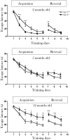
Age-dependent deficit in spatial learning in Cnr1−/− mice. Curves show the escape latencies during the acquisition and reversal trials. *P < 0.05, **P < 0.01, ***P < 0.001 difference between the genotypes (two-way ANOVA followed by Bonferroni's t test; n = 12 per group).
Fig. 3.
Fig. 3.
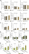
Neuronal loss and enhanced neuroinflammation in the hippocampus of GABA-Cnr1−/− but not in Glu-Cnr1−/−. (A) Quantitative stereological analysis revealed a decrease in the number of principal neurons in the hippocampus of 12-mo-old GABA-Cnr1−/− mice. (B) Total number of Iba1-positive cells in the CA1 region of the hippocampus is increased in 12-mo-old GABA-Cnr1−/− but not in Glu-Cnr1−/− animals. (C) The age-dependent increase in the fraction of GFAP-immunopositive areas in the CA1 and CA3 regions of the hippocampus is more pronounced in 12-mo-old GABA-Cnr1−/− mice than their wild-type littermates. (*P < 0.05, **P < 0.01 Student's t test; n = 3 mice/group) (D) Comparison of 2- and 12-mo-old animals revealed a similar expression of cytokines in Glu-Cnr1−/− mice and in their control littermates. (E) In GABA-Cnr1−/− animals, IL-6 and TNF but not IL-1β expression significantly increased in aging. In their wild-type littermates there was no age-related change in the expression levels of cytokines.

References

    1. Murakami S, Murakami H. The effects of aging and oxidative stress on learning behavior in C. elegans. Neurobiol Aging. 2005;26:899–905. - PubMed
    1. Li SC, et al. Transformations in the couplings among intellectual abilities and constituent cognitive processes across the life span. Psychol Sci. 2004;15:155–163. - PubMed
    1. Kirkwood TB, Franceschi C. Is aging as complex as it would appear? New perspectives in aging research. Ann N Y Acad Sci. 1992;663:412–417. - PubMed
    1. Lucin KM, Wyss-Coray T. Immune activation in brain aging and neurodegeneration: Too much or too little? Neuron. 2009;64:110–122. - PMC - PubMed
    1. Perry VH, Nicoll JA, Holmes C. Microglia in neurodegenerative disease. Nat Rev Neurol. 2010;6:193–201. - PubMed

Publication types

MeSH terms

LinkOut - more resources