. 2011 Jul 5;108(27):11193-8.
doi: 10.1073/pnas.1019536108. Epub 2011 Jun 20.

MicroRNA-155 as a proinflammatory regulator in clinical and experimental arthritis

Affiliations

MicroRNA-155 as a proinflammatory regulator in clinical and experimental arthritis

Mariola Kurowska-Stolarska et al. Proc Natl Acad Sci U S A. .

Abstract

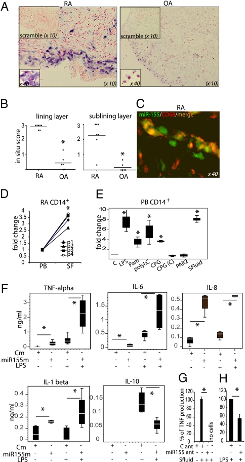
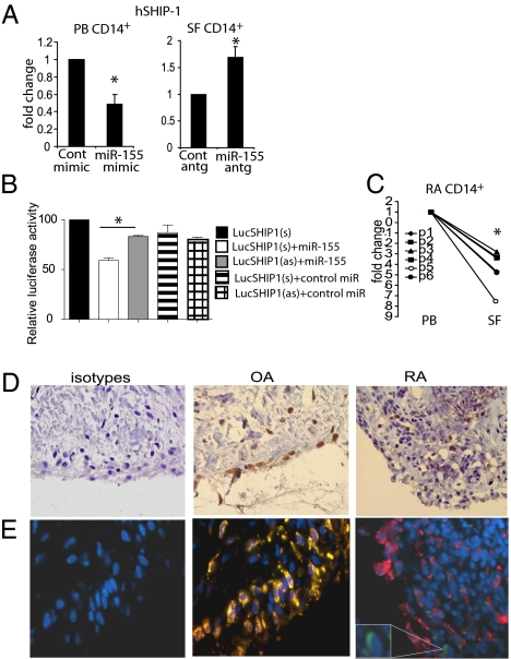
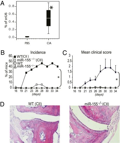
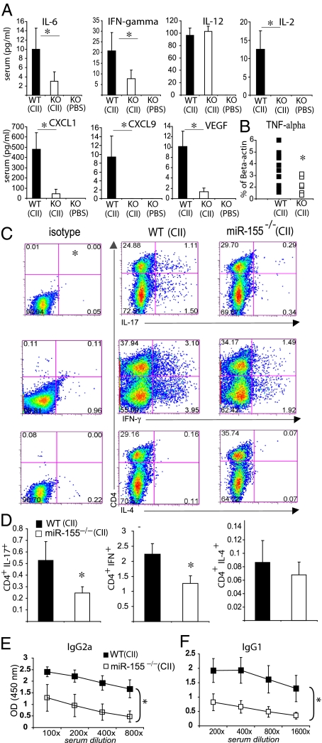
MicroRNA (miRNA) species (miR) regulate mRNA translation and are implicated as mediators of disease pathology via coordinated regulation of molecular effector pathways. Unraveling miR disease-related activities will facilitate future therapeutic interventions. miR-155 recently has been identified with critical immune regulatory functions. Although detected in articular tissues, the functional role of miR-155 in inflammatory arthritis has not been defined. We report here that miR-155 is up-regulated in synovial membrane and synovial fluid (SF) macrophages from patients with rheumatoid arthritis (RA). The increased expression of miR-155 in SF CD14(+) cells was associated with lower expression of the miR-155 target, Src homology 2-containing inositol phosphatase-1 (SHIP-1), an inhibitor of inflammation. Similarly, SHIP-1 expression was decreased in CD68(+) cells in the synovial lining layer in RA patients as compared with osteoarthritis patients. Overexpression of miR-155 in PB CD14(+) cells led to down-regulation of SHIP-1 and an increase in the production of proinflammatory cytokines. Conversely, inhibition of miR-155 in RA synovial CD14(+) cells reduced TNF-α production. Finally, miR-155-deficient mice are resistant to collagen-induced arthritis, with profound suppression of antigen-specific Th17 cell and autoantibody responses and markedly reduced articular inflammation. Our data therefore identify a role of miR-155 in clinical and experimental arthritis and suggest that miR-155 may be an intriguing therapeutic target.


Conflict of interest statement

The authors declare no conflict of interest.

Figures

Fig. 1.
Fig. 1.
MiR-155 is up-regulated in RA synovial macrophages and monocytes and promotes production of proinflammatory cytokines. (A and B) Expression of miR-155 in RA and OA biopsies. (A) Representative staining. (B) Quantitative evaluation of miR-155 expression, *P < 0.05 RA vs. OA. (C) Representative photograph of double staining for miR-155 (green) and macrophage marker CD68 (red) in RA lining layer. Double-positive cells are orange or yellow. (D) Expression of miR-155 in paired PB and SF CD14+ cells from RA patients (n = 4), *P < 0.05 PB vs. SF. (E) TLR ligands and RA SF up-regulate miR-155 expression in PB CD14+ cells (n = 3). Cells were stimulated with TLR ligands or PAR2 agonist or 25% SF for 24 h as described in Materials and Methods. *P < 0.05 stimulated vs nonstimulated. (F) PB CD14+ cells overexpressing miR-155 produce proinflammatory cytokines. CD14+ cells were transfected with control or miR-155 mimic (20 nM). After 24 h, LPS (100 ng/mL) was added to some wells for a further 16 h (n = 5). (G and H) PB CD14+ cells and RA SF CD14+ cells (n = 2 and 3, respectively) were transfected with control or miR-155 antagomir. After 24 h cells were exposed to SF (F) or LPS (G) for a further 24 h. *P < 0.05 as indicated. Data are means ± SEM or box-and-whisker diagrams. Cm, control mimic; miR155ant, miR-155 antagomir; miR155m, miR-155 mimic, C ant, control antagomir; PAR2, PAR2 agonist, (SLIGKV-NH2). The in situ scoring system is described in SI Materials and Methods.
Fig. 2.
Fig. 2.
The miR-155 target SHIP-1 is down-regulated in RA SF CD14+ cells and in RA synovial tissue macrophages. (A) miR-155 regulates expression of SHIP-1 in RA PB and SF CD14+ cells. Cells were transfected with miR-155 mimic or antagomir or with appropriate controls as described in Fig. 1. Quantitative analysis of SHIP-1 mRNA expression is shown (n = 3). (B) miR-155 binds directly to the 3′ UTR of human SHIP-1 mRNA. pMir-hSHIP1 (sense) and pMir-hSHIP1 (antisense) 3′ UTR luciferase plasmids were cotransfected with control or miR-155 mimic (40 nM) in HEK293 cells. Luciferase activity was analyzed at 24 h. (C) Expression of SHIP-1 in paired PB and SF CD14+ cells from RA patients (n = 6). Data are means ± SEM. *P < 0.05. (D and E) Synovial specimens from RA (n = 5) and OA (n = 5) patients stained with anti-human SHIP-1 (D, brown) and counterstained with anti-CD68 antibody (E). Representative staining of one of five specimens is shown; blue, DAPI; red, CD68+; green, SHIP-1+; yellow, CD68+/SHIP-1+. (Magnification: 63×.) Enlargements in E show a SHIP-1+ cell (green). Cont, control mimic; hSHIP-1, human SHIP-1; miR155antg, miR-155 antagomir; miR155m, miR-155 mimic; Cont antg, control antagomir. Relative luciferase activity is the maximal activity in the presence of pMiR plasmid alone.
Fig. 3.
Fig. 3.
miR-155 is crucial for the development of CIA. On day 0, miR-155−/− and WT mice (n = 15–17 per group) were injected intradermally with either a type II chicken collagen/Freund's complete adjuvant emulsion (CII/CFA) (200 μg) or PBS. On day 21, type II chicken collagen in PBS (200 μg) was injected i.p. Mice were killed on day 34. (A) miR-155 is up-regulated in the articular tissue of WT mice with CIA. (B and C) The incidence (B) and mean clinical score (C) are shown. In contrast to WT littermates, miR-155−/− mice do not develop signs of CIA. (D) In contrast to WT mice, miR-155−/− mice did not develop articular inflammation and degradation of cartilage and bone. (Magnification: ×10.) Data are means ± SEM or box-and-whisker diagrams. (n = 15–17); *P < 0.05, miR-155−/− vs. WT mice (CIA protocol).
Fig. 4.
Fig. 4.
miR-155−/− mice exhibit reduced development of T- and B-cell responses during CIA. Groups are as in Fig. 3. CII/miR-155−/− mice show (A) reduced systemic cytokine and chemokine concentrations and (B) reduced expression of TNF-α mRNA in articular tissue. (C and D) CII-miR-155−/− mice show a reduced differentiation of Th17 and Th1 cells in DLN. Representative staining (C) and quantitative evaluation (D) are shown. (E and F) miR-155 is required for the production of collagen-specific antibodies. Quantitative evaluation of antibody activity in isotypes IgG2a (E) and IgG1 (F) is shown. Data are means ± SEM, n = 15–17; *P < 0.05, miR-155−/− vs. WT mice (CIA protocol). CII, type II chicken collagen.

Comment in

References

    1. McInnes IB, Schett G. Cytokines in the pathogenesis of rheumatoid arthritis. Nat Rev Immunol. 2007;7:429–442. - PubMed
    1. Bartel DP. MicroRNAs: Genomics, biogenesis, mechanism, and function. Cell. 2004;116:281–297. - PubMed
    1. O'Connell RM, Rao DS, Chaudhuri AA, Baltimore D. Physiological and pathological roles for microRNAs in the immune system. Nat Rev Immunol. 2010;10:111–122. - PubMed
    1. Pillai RS, Bhattacharyya SN, Filipowicz W. Repression of protein synthesis by miRNAs: How many mechanisms? Trends Cell Biol. 2007;17:118–126. - PubMed
    1. Xiao C, et al. Lymphoproliferative disease and autoimmunity in mice with increased miR-17-92 expression in lymphocytes. Nat Immunol. 2008;9:405–414. - PMC - PubMed

Publication types

MeSH terms